 Movie
Movie Controller


+ Open data

- Basic information
| Entry | Database: PDB / ID: 4k1n | ||||||
|---|---|---|---|---|---|---|---|
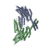
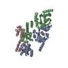
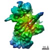
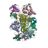
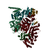
| Title | Crystal structure of full-length mouse alphaE-catenin | ||||||
 Components Components | Catenin alpha-1 | ||||||
 Keywords Keywords | CELL ADHESION / four-helix bundle / beta-catenin / F-actin | ||||||
| Function / homology |  Function and homology information Function and homology informationnegative regulation of integrin-mediated signaling pathway / VEGFR2 mediated vascular permeability / RHO GTPases activate IQGAPs / Adherens junctions interactions / gap junction assembly / epithelial cell-cell adhesion / zonula adherens / gamma-catenin binding / cellular response to indole-3-methanol / vinculin binding ...negative regulation of integrin-mediated signaling pathway / VEGFR2 mediated vascular permeability / RHO GTPases activate IQGAPs / Adherens junctions interactions / gap junction assembly / epithelial cell-cell adhesion / zonula adherens / gamma-catenin binding / cellular response to indole-3-methanol / vinculin binding / flotillin complex / negative regulation of cell motility / Myogenesis / apical junction assembly / positive regulation of extrinsic apoptotic signaling pathway in absence of ligand / positive regulation of smoothened signaling pathway / catenin complex / negative regulation of protein localization to nucleus / axon regeneration / negative regulation of neuroblast proliferation / smoothened signaling pathway / establishment or maintenance of cell polarity / odontogenesis of dentin-containing tooth / neuroblast proliferation / negative regulation of extrinsic apoptotic signaling pathway in absence of ligand / intercalated disc / ovarian follicle development / extrinsic apoptotic signaling pathway in absence of ligand / acrosomal vesicle / cell motility / integrin-mediated signaling pathway / adherens junction / protein localization / cell-cell adhesion / beta-catenin binding / response to estrogen / male gonad development / cell migration / actin filament binding / cell-cell junction / actin cytoskeleton / cell junction / lamellipodium / regulation of cell population proliferation / cadherin binding / intracellular membrane-bounded organelle / apoptotic process / protein-containing complex binding / negative regulation of apoptotic process / structural molecule activity / identical protein binding / plasma membraneSimilarity search - Function | ||||||
| Biological species |  Mus musculus (house mouse) Mus musculus (house mouse) | ||||||
| Method | X-RAY DIFFRACTION / SYNCHROTRON / MOLECULAR REPLACEMENT / Resolution: 6.5 Å | ||||||
 Authors Authors | Ishiyama, N. / Ikura, M. | ||||||
 Citation Citation | Journal: J.Biol.Chem. / Year: 2013 Title: An autoinhibited structure of alpha-catenin and its implications for vinculin recruitment to adherens junctions. Authors: Ishiyama, N. / Tanaka, N. / Abe, K. / Yang, Y.J. / Abbas, Y.M. / Umitsu, M. / Nagar, B. / Bueler, S.A. / Rubinstein, J.L. / Takeichi, M. / Ikura, M. | ||||||
| History |
|
- Structure visualization
| Structure viewer | Molecule: MolmilJmol/JSmol |
|---|
- Downloads & links
-Download
| PDBx/mmCIF format | 4k1n.cif.gz | 196.3 KB | Display | PDBx/mmCIF format |
|---|---|---|---|---|
| PDB format | pdb4k1n.ent.gz | 143.6 KB | Display | PDB format |
| PDBx/mmJSON format | 4k1n.json.gz | Tree view | PDBx/mmJSON format | |
| Others |  Other downloads Other downloads |
-Validation report
| Arichive directory | https://data.pdbj.org/pub/pdb/validation_reports/k1/4k1nftp://data.pdbj.org/pub/pdb/validation_reports/k1/4k1n | HTTPS FTP |
|---|
-Related structure data
| Related structure data | 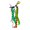 4k1oC 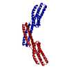 1dovS 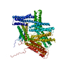 1st6S C: citing same article ( S: Starting model for refinement |
|---|---|
| Similar structure data |


-Links
 PDBj
PDBj

- Assembly
| Deposited unit | 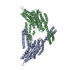
| ||||||||
|---|---|---|---|---|---|---|---|---|---|
| 1 |
| ||||||||
| Unit cell |
|
-Components
| #1: Protein | / 102 kDa cadherin-associated protein / Alpha E-catenin / CAP102 Mass: 100812.016 Da / Num. of mol.: 2 Source method: isolated from a genetically manipulated source Source: (gene. exp.) Mus musculus (house mouse) / Gene: Catna1, Ctnna1 / Plasmid: pET28a / Production host:  Escherichia coli (E. coli) / Strain (production host): BL21-CodonPlus / References: UniProt: P26231 Escherichia coli (E. coli) / Strain (production host): BL21-CodonPlus / References: UniProt: P26231 |
|---|
-Experimental details
-Experiment
| Experiment | Method: X-RAY DIFFRACTION / Number of used crystals: 1 |
|---|
- Sample preparation
| Crystal | Density Matthews: 4.1 Å3/Da / Density % sol: 69.99 % |
|---|---|
| Crystal grow | Temperature: 293 K / Method: vapor diffusion Details: 0.2 M Sodium Citrate, 20% (w/v) PEG-3350, vapor diffusion, temperature 293K |
-Data collection
| Diffraction |
| |||||||||||||||||||||||||||||||||||||||||||||||||||||||||||||||||||||||||||||||||||||||||||||||||||||||||||||||||||||||||||||||||||||||||||||||||||
|---|---|---|---|---|---|---|---|---|---|---|---|---|---|---|---|---|---|---|---|---|---|---|---|---|---|---|---|---|---|---|---|---|---|---|---|---|---|---|---|---|---|---|---|---|---|---|---|---|---|---|---|---|---|---|---|---|---|---|---|---|---|---|---|---|---|---|---|---|---|---|---|---|---|---|---|---|---|---|---|---|---|---|---|---|---|---|---|---|---|---|---|---|---|---|---|---|---|---|---|---|---|---|---|---|---|---|---|---|---|---|---|---|---|---|---|---|---|---|---|---|---|---|---|---|---|---|---|---|---|---|---|---|---|---|---|---|---|---|---|---|---|---|---|---|---|---|---|---|
| Diffraction source | Source: SYNCHROTRON / Site: APS  / Beamline: 19-ID / Wavelength: 0.97918 Å / Beamline: 19-ID / Wavelength: 0.97918 Å | |||||||||||||||||||||||||||||||||||||||||||||||||||||||||||||||||||||||||||||||||||||||||||||||||||||||||||||||||||||||||||||||||||||||||||||||||||
| Detector | Type: SBC-3 / Detector: CCD / Date: Apr 15, 2007 | |||||||||||||||||||||||||||||||||||||||||||||||||||||||||||||||||||||||||||||||||||||||||||||||||||||||||||||||||||||||||||||||||||||||||||||||||||
| Radiation | Protocol: SINGLE WAVELENGTH / Monochromatic (M) / Laue (L): M / Scattering type: x-ray | |||||||||||||||||||||||||||||||||||||||||||||||||||||||||||||||||||||||||||||||||||||||||||||||||||||||||||||||||||||||||||||||||||||||||||||||||||
| Radiation wavelength | Wavelength: 0.97918 Å / Relative weight: 1 | |||||||||||||||||||||||||||||||||||||||||||||||||||||||||||||||||||||||||||||||||||||||||||||||||||||||||||||||||||||||||||||||||||||||||||||||||||
| Reflection | Resolution: 6.5→50 Å / Num. obs: 6581 / % possible obs: 99.7 % / Observed criterion σ(F): -3 / Observed criterion σ(I): 0 / Redundancy: 5.2 % / Rsym value: 0.128 / Χ2: 1.044 / Net I/σ(I): 14.3 | |||||||||||||||||||||||||||||||||||||||||||||||||||||||||||||||||||||||||||||||||||||||||||||||||||||||||||||||||||||||||||||||||||||||||||||||||||
| Reflection shell |
|
- Processing
| Software |
| |||||||||||||||||||||||||||||||||||||||||||||||||||||||||||||||||||||||||||||
|---|---|---|---|---|---|---|---|---|---|---|---|---|---|---|---|---|---|---|---|---|---|---|---|---|---|---|---|---|---|---|---|---|---|---|---|---|---|---|---|---|---|---|---|---|---|---|---|---|---|---|---|---|---|---|---|---|---|---|---|---|---|---|---|---|---|---|---|---|---|---|---|---|---|---|---|---|---|---|
| Refinement | Method to determine structure: MOLECULAR REPLACEMENT Starting model: 1DOV and 1ST6 Resolution: 6.5→50 Å / Occupancy max: 1 / Occupancy min: 1 / σ(F): 0 / Stereochemistry target values: Engh & Huber
| |||||||||||||||||||||||||||||||||||||||||||||||||||||||||||||||||||||||||||||
| Solvent computation | Bsol: 333.391 Å2 | |||||||||||||||||||||||||||||||||||||||||||||||||||||||||||||||||||||||||||||
| Displacement parameters | Biso max: 354.92 Å2 / Biso mean: 354.92 Å2 / Biso min: 354.92 Å2
| |||||||||||||||||||||||||||||||||||||||||||||||||||||||||||||||||||||||||||||
| Refinement step | Cycle: LAST / Resolution: 6.5→50 Å
| |||||||||||||||||||||||||||||||||||||||||||||||||||||||||||||||||||||||||||||
| Refine LS restraints |
| |||||||||||||||||||||||||||||||||||||||||||||||||||||||||||||||||||||||||||||
| LS refinement shell | Refine-ID: X-RAY DIFFRACTION / Total num. of bins used: 10
| |||||||||||||||||||||||||||||||||||||||||||||||||||||||||||||||||||||||||||||
| Xplor file |
|
