 Movie
Movie Controller

[English] 日本語
 Yorodumi
- PDB-4jfs: Crystal structure of a bacterial fucosidase with iminosugar inhib... -
+ Open data

- Basic information
| Entry | Database: PDB / ID: 4jfs | ||||||
|---|---|---|---|---|---|---|---|
| Title | Crystal structure of a bacterial fucosidase with iminosugar inhibitor 4-epi-(+)-Codonopsinine | ||||||
 Components Components | alpha-L-fucosidase | ||||||
 Keywords Keywords | HYDROLASE/HYDROLASE INHIBITOR / alpha-L-fucosidase / HYDROLASE-HYDROLASE INHIBITOR complex | ||||||
| Function / homology |  Function and homology informationalpha-L-fucosidase activity / fucose metabolic process / glycoside catabolic process / lysosome Function and homology informationalpha-L-fucosidase activity / fucose metabolic process / glycoside catabolic process / lysosomeSimilarity search - Function | ||||||
| Biological species |  Bacteroides thetaiotaomicron (bacteria) Bacteroides thetaiotaomicron (bacteria) | ||||||
| Method | X-RAY DIFFRACTION / SYNCHROTRON / MOLECULAR REPLACEMENT / Resolution: 2 Å | ||||||
 Authors Authors | Wright, D.W. / Davies, G.J. | ||||||
 Citation Citation | Journal: Chemistry / Year: 2013 Title: alpha-L-fucosidase inhibition by pyrrolidine-ferrocene hybrids: rationalization of ligand-binding properties by structural studies. Authors: Hottin, A. / Wright, D.W. / Steenackers, A. / Delannoy, P. / Dubar, F. / Biot, C. / Davies, G.J. / Behr, J.B. | ||||||
| History |
|
- Structure visualization
| Structure viewer | Molecule: MolmilJmol/JSmol |
|---|
- Downloads & links
-Download
| PDBx/mmCIF format | 4jfs.cif.gz | 211.5 KB | Display | PDBx/mmCIF format |
|---|---|---|---|---|
| PDB format | pdb4jfs.ent.gz | 164.1 KB | Display | PDB format |
| PDBx/mmJSON format | 4jfs.json.gz | Tree view | PDBx/mmJSON format | |
| Others |  Other downloads Other downloads |
-Validation report
| Arichive directory | https://data.pdbj.org/pub/pdb/validation_reports/jf/4jfsftp://data.pdbj.org/pub/pdb/validation_reports/jf/4jfs | HTTPS FTP |
|---|
-Related structure data
| Related structure data |  4jftC  4jfuC  4jfvC  4jfwC 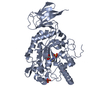 4j27S S: Starting model for refinement C: citing same article ( |
|---|---|
| Similar structure data |


-Links
 PDBj
PDBj


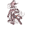
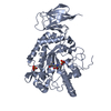
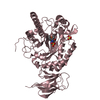
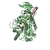
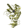
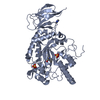
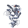
- Assembly
| Deposited unit | 
| ||||||||||||||||||
|---|---|---|---|---|---|---|---|---|---|---|---|---|---|---|---|---|---|---|---|
| 1 | 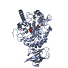
| ||||||||||||||||||
| 2 | 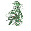
| ||||||||||||||||||
| Unit cell |
| ||||||||||||||||||
| Noncrystallographic symmetry (NCS) | NCS domain:
NCS domain segments: Component-ID: 0 / Ens-ID: 1 / Beg auth comp-ID: GLU / Beg label comp-ID: GLU / End auth comp-ID: ARG / End label comp-ID: ARG / Refine code: 0 / Auth seq-ID: 35 - 470 / Label seq-ID: 1 - 436
|
-Components
-Protein , 1 types, 2 molecules AB
| #1: Protein | Mass: 52080.930 Da / Num. of mol.: 2 / Fragment: RESIDUES 35-484 Source method: isolated from a genetically manipulated source Source: (gene. exp.) Bacteroides thetaiotaomicron (bacteria)Strain: VPI-5482 / Plasmid: pET-YSBLIC / Production host: Escherichia coli (E. coli) / Strain (production host): BL21(DE3) / References: UniProt: Q8A3I4 |
|---|
-Non-polymers , 6 types, 865 molecules 


| #2: Chemical |  Mass: 237.295 Da / Num. of mol.: 2 / Source method: obtained synthetically / Formula: C13H19NO3 Mass: 237.295 Da / Num. of mol.: 2 / Source method: obtained synthetically / Formula: C13H19NO3#3: Chemical | ChemComp-IMD / Imidazole Mass: 69.085 Da / Num. of mol.: 5 / Source method: obtained synthetically / Formula: C3H5N2 Mass: 69.085 Da / Num. of mol.: 5 / Source method: obtained synthetically / Formula: C3H5N2#4: Chemical | ChemComp-SO4 / Sulfate Mass: 96.063 Da / Num. of mol.: 4 / Source method: obtained synthetically / Formula: SO4 Mass: 96.063 Da / Num. of mol.: 4 / Source method: obtained synthetically / Formula: SO4#5: Chemical | ChemComp-GOL / | Glycerol Mass: 92.094 Da / Num. of mol.: 1 / Source method: obtained synthetically / Formula: C3H8O3 Mass: 92.094 Da / Num. of mol.: 1 / Source method: obtained synthetically / Formula: C3H8O3#6: Chemical | ChemComp-EPE / | HEPES Mass: 238.305 Da / Num. of mol.: 1 / Source method: obtained synthetically / Formula: C8H18N2O4S / Comment: pH buffer*YM Mass: 238.305 Da / Num. of mol.: 1 / Source method: obtained synthetically / Formula: C8H18N2O4S / Comment: pH buffer*YM#7: Water | ChemComp-HOH / | WaterMass: 18.015 Da / Num. of mol.: 852 / Source method: isolated from a natural source / Formula: H2O |
|---|
-Experimental details
-Experiment
| Experiment | Method: X-RAY DIFFRACTION / Number of used crystals: 1 |
|---|
- Sample preparation
| Crystal | Density Matthews: 3.05 Å3/Da / Density % sol: 59.73 % |
|---|---|
| Crystal grow | Temperature: 291.5 K / Method: vapor diffusion, hanging drop / pH: 7 Details: 0.13 M ammonium sulfate, 12% PEG 6K, 0.1M imidazole pH 7, VAPOR DIFFUSION, HANGING DROP, temperature 291.5K |
-Data collection
| Diffraction | Mean temperature: 100 K | ||||||||||||||||||||||||||||||||||||||||||||||||||||||||||||||||||||||||||||||||||||||||
|---|---|---|---|---|---|---|---|---|---|---|---|---|---|---|---|---|---|---|---|---|---|---|---|---|---|---|---|---|---|---|---|---|---|---|---|---|---|---|---|---|---|---|---|---|---|---|---|---|---|---|---|---|---|---|---|---|---|---|---|---|---|---|---|---|---|---|---|---|---|---|---|---|---|---|---|---|---|---|---|---|---|---|---|---|---|---|---|---|---|
| Diffraction source | Source: SYNCHROTRON / Site: Diamond  / Beamline: I04-1 / Wavelength: 0.9173 Å / Beamline: I04-1 / Wavelength: 0.9173 Å | ||||||||||||||||||||||||||||||||||||||||||||||||||||||||||||||||||||||||||||||||||||||||
| Detector | Type: PILATUS 2M / Detector: PIXEL / Date: Mar 8, 2012 | ||||||||||||||||||||||||||||||||||||||||||||||||||||||||||||||||||||||||||||||||||||||||
| Radiation | Monochromator: Si 111 CHANNEL / Protocol: SINGLE WAVELENGTH / Monochromatic (M) / Laue (L): M / Scattering type: x-ray | ||||||||||||||||||||||||||||||||||||||||||||||||||||||||||||||||||||||||||||||||||||||||
| Radiation wavelength | Wavelength: 0.9173 Å / Relative weight: 1 | ||||||||||||||||||||||||||||||||||||||||||||||||||||||||||||||||||||||||||||||||||||||||
| Reflection | Resolution: 1.578→97.101 Å / Num. all: 83920 / Num. obs: 83920 / % possible obs: 99.2 % / Observed criterion σ(F): 0 / Observed criterion σ(I): 0 / Redundancy: 3.9 % / Rsym value: 0.099 / Net I/σ(I): 9.2 | ||||||||||||||||||||||||||||||||||||||||||||||||||||||||||||||||||||||||||||||||||||||||
| Reflection shell | Diffraction-ID: 1
|
- Processing
| Software |
| |||||||||||||||||||||||||||||||||||||||||||||||||||||||||||||||||||||||||||
|---|---|---|---|---|---|---|---|---|---|---|---|---|---|---|---|---|---|---|---|---|---|---|---|---|---|---|---|---|---|---|---|---|---|---|---|---|---|---|---|---|---|---|---|---|---|---|---|---|---|---|---|---|---|---|---|---|---|---|---|---|---|---|---|---|---|---|---|---|---|---|---|---|---|---|---|---|
| Refinement | Method to determine structure: MOLECULAR REPLACEMENT Starting model: 4J27 Resolution: 2→97.1 Å / Cor.coef. Fo:Fc: 0.964 / Cor.coef. Fo:Fc free: 0.945 / WRfactor Rfree: 0.185 / WRfactor Rwork: 0.1503 / Occupancy max: 1 / Occupancy min: 0.5 / FOM work R set: 0.8785 / SU B: 3.416 / SU ML: 0.092 / SU R Cruickshank DPI: 0.1316 / SU Rfree: 0.1246 / Cross valid method: THROUGHOUT / σ(F): 0 / ESU R: 0.132 / ESU R Free: 0.125 / Stereochemistry target values: MAXIMUM LIKELIHOOD Details: HYDROGENS HAVE BEEN ADDED IN THE RIDING POSITIONS U VALUES: REFINED INDIVIDUALLY. THE COORDINATES FROM 4J27 WERE CONVERTED INTO THE (ALMOST) ISOMORPHOUS CELL OF 4JFS TO BUILD A STARTING ...Details: HYDROGENS HAVE BEEN ADDED IN THE RIDING POSITIONS U VALUES: REFINED INDIVIDUALLY. THE COORDINATES FROM 4J27 WERE CONVERTED INTO THE (ALMOST) ISOMORPHOUS CELL OF 4JFS TO BUILD A STARTING MODEL DIRECTLY. THE RFREE FLAG FROM 4J27 WAS USED FOR THIS DATASET.
| |||||||||||||||||||||||||||||||||||||||||||||||||||||||||||||||||||||||||||
| Solvent computation | Ion probe radii: 0.8 Å / Shrinkage radii: 0.8 Å / VDW probe radii: 1.2 Å / Solvent model: MASK | |||||||||||||||||||||||||||||||||||||||||||||||||||||||||||||||||||||||||||
| Displacement parameters | Biso max: 86 Å2 / Biso mean: 25.8593 Å2 / Biso min: 13.11 Å2
| |||||||||||||||||||||||||||||||||||||||||||||||||||||||||||||||||||||||||||
| Refinement step | Cycle: LAST / Resolution: 2→97.1 Å
| |||||||||||||||||||||||||||||||||||||||||||||||||||||||||||||||||||||||||||
| Refine LS restraints |
| |||||||||||||||||||||||||||||||||||||||||||||||||||||||||||||||||||||||||||
| Refine LS restraints NCS | Ens-ID: 1 / Number: 26798 / Refine-ID: X-RAY DIFFRACTION / Type: interatomic distance / Rms dev position: 0.07 Å / Weight position: 0.05
| |||||||||||||||||||||||||||||||||||||||||||||||||||||||||||||||||||||||||||
| LS refinement shell | Resolution: 2→2.052 Å / Total num. of bins used: 20
|
