 Movie
Movie Controller

[English] 日本語
 Yorodumi
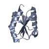
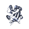
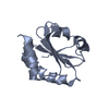
- PDB-4gwr: Crystal Structure of the second catalytic domain of protein disul... -
+ Open data

- Basic information
| Entry | Database: PDB / ID: 4gwr | ||||||
|---|---|---|---|---|---|---|---|
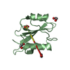
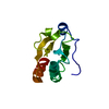
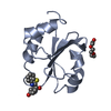
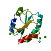
| Title | Crystal Structure of the second catalytic domain of protein disulfide isomerase P5 | ||||||
 Components Components | Protein disulfide-isomerase A6 | ||||||
 Keywords Keywords |  ISOMERASE / thioredoxin-like fold / disulfide isomerase / BiP / endoplasmic reticulum ISOMERASE / thioredoxin-like fold / disulfide isomerase / BiP / endoplasmic reticulum | ||||||
| Function / homology |  Function and homology informationprotein disulfide-isomerase / endoplasmic reticulum chaperone complex / XBP1(S) activates chaperone genes / protein disulfide isomerase activity / endoplasmic reticulum-Golgi intermediate compartment / protein-disulfide reductase activity / response to endoplasmic reticulum stress / Post-translational protein phosphorylation / Regulation of Insulin-like Growth Factor (IGF) transport and uptake by Insulin-like Growth Factor Binding Proteins (IGFBPs) / melanosome ...protein disulfide-isomerase / endoplasmic reticulum chaperone complex / XBP1(S) activates chaperone genes / protein disulfide isomerase activity / endoplasmic reticulum-Golgi intermediate compartment / protein-disulfide reductase activity / response to endoplasmic reticulum stress / Post-translational protein phosphorylation / Regulation of Insulin-like Growth Factor (IGF) transport and uptake by Insulin-like Growth Factor Binding Proteins (IGFBPs) / melanosome / protein folding / endoplasmic reticulum lumen / endoplasmic reticulum membrane / endoplasmic reticulum / extracellular space / extracellular exosome / plasma membrane / cytosol Function and homology informationprotein disulfide-isomerase / endoplasmic reticulum chaperone complex / XBP1(S) activates chaperone genes / protein disulfide isomerase activity / endoplasmic reticulum-Golgi intermediate compartment / protein-disulfide reductase activity / response to endoplasmic reticulum stress / Post-translational protein phosphorylation / Regulation of Insulin-like Growth Factor (IGF) transport and uptake by Insulin-like Growth Factor Binding Proteins (IGFBPs) / melanosome ...protein disulfide-isomerase / endoplasmic reticulum chaperone complex / XBP1(S) activates chaperone genes / protein disulfide isomerase activity / endoplasmic reticulum-Golgi intermediate compartment / protein-disulfide reductase activity / response to endoplasmic reticulum stress / Post-translational protein phosphorylation / Regulation of Insulin-like Growth Factor (IGF) transport and uptake by Insulin-like Growth Factor Binding Proteins (IGFBPs) / melanosome / protein folding / endoplasmic reticulum lumen / endoplasmic reticulum membrane / endoplasmic reticulum / extracellular space / extracellular exosome / plasma membrane / cytosolSimilarity search - Function | ||||||
| Biological species |  Homo sapiens (human) Homo sapiens (human) | ||||||
| Method | X-RAY DIFFRACTION / SYNCHROTRON / MOLECULAR REPLACEMENT / Resolution: 1.81 Å | ||||||
 Authors Authors | Vinaik, R. / Kozlov, G. / Gehring, K. | ||||||
 Citation Citation | Journal: To be Published / Year: 2013 Title: To be published Authors: Vinaik, R. / Kozlov, G. / Gehring, K. | ||||||
| History |
|
- Structure visualization
| Structure viewer | Molecule: MolmilJmol/JSmol |
|---|
- Downloads & links
-Download
| PDBx/mmCIF format | 4gwr.cif.gz | 57.2 KB | Display | PDBx/mmCIF format |
|---|---|---|---|---|
| PDB format | pdb4gwr.ent.gz | 41.1 KB | Display | PDB format |
| PDBx/mmJSON format | 4gwr.json.gz | Tree view | PDBx/mmJSON format | |
| Others |  Other downloads Other downloads |
-Validation report
| Arichive directory | https://data.pdbj.org/pub/pdb/validation_reports/gw/4gwrftp://data.pdbj.org/pub/pdb/validation_reports/gw/4gwr | HTTPS FTP |
|---|
-Related structure data
| Related structure data |  4ef0S S: Starting model for refinement |
|---|---|
| Similar structure data |


-Links
 PDBj
PDBj


- Assembly
| Deposited unit | 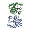
| ||||||||
|---|---|---|---|---|---|---|---|---|---|
| 1 | 
| ||||||||
| 2 | 
| ||||||||
| Unit cell |
|
-Components
| #1: Protein | Mass: 13889.551 Da / Num. of mol.: 2 / Fragment: second catalytic domain, UNP residues 160-274 Source method: isolated from a genetically manipulated source Source: (gene. exp.) Homo sapiens (human) / Gene: PDIA6, ERP5, P5, TXNDC7 / Plasmid: pET23 / Production host:  Escherichia coli (E. coli) / Strain (production host): BL21 / References: UniProt: Q15084, protein disulfide-isomerase Escherichia coli (E. coli) / Strain (production host): BL21 / References: UniProt: Q15084, protein disulfide-isomerase#2: Water | ChemComp-HOH / | Water Mass: 18.015 Da / Num. of mol.: 89 / Source method: isolated from a natural source / Formula: H2O Mass: 18.015 Da / Num. of mol.: 89 / Source method: isolated from a natural source / Formula: H2O |
|---|
-Experimental details
-Experiment
| Experiment | Method: X-RAY DIFFRACTION / Number of used crystals: 1 |
|---|
- Sample preparation
| Crystal | Density Matthews: 1.89 Å3/Da / Density % sol: 34.81 % |
|---|---|
| Crystal grow | Temperature: 294 K / Method: vapor diffusion, hanging drop / pH: 3.5 Details: 0.1M citric acid, 25% PEG3350, pH 3.5, VAPOR DIFFUSION, HANGING DROP, temperature 294K |
-Data collection
| Diffraction | Mean temperature: 100 K |
|---|---|
| Diffraction source | Source: SYNCHROTRON / Site: CHESS  / Beamline: A1 / Wavelength: 0.976 Å / Beamline: A1 / Wavelength: 0.976 Å |
| Detector | Type: ADSC QUANTUM 210 / Detector: CCD / Date: Jun 10, 2012 |
| Radiation | Monochromator: Si 111 CHANNEL / Protocol: SINGLE WAVELENGTH / Monochromatic (M) / Laue (L): M / Scattering type: x-ray |
| Radiation wavelength | Wavelength: 0.976 Å / Relative weight: 1 |
| Reflection | Resolution: 1.8→50 Å / Num. all: 17641 / Num. obs: 17069 / % possible obs: 96.76 % / Observed criterion σ(F): 1 / Observed criterion σ(I): 1 / Redundancy: 2.4 % / Rsym value: 0.061 / Net I/σ(I): 15.8 |
| Reflection shell | Resolution: 1.8→1.83 Å / Redundancy: 1.9 % / Mean I/σ(I) obs: 2.5 / Num. unique all: 1136 / Rsym value: 0.268 / % possible all: 86.01 |
- Processing
| Software |
| ||||||||||||||||||||||||||||||||||||||||||||||||||||||||||||||||||||||||||||||||||||||||||
|---|---|---|---|---|---|---|---|---|---|---|---|---|---|---|---|---|---|---|---|---|---|---|---|---|---|---|---|---|---|---|---|---|---|---|---|---|---|---|---|---|---|---|---|---|---|---|---|---|---|---|---|---|---|---|---|---|---|---|---|---|---|---|---|---|---|---|---|---|---|---|---|---|---|---|---|---|---|---|---|---|---|---|---|---|---|---|---|---|---|---|---|
| Refinement | Method to determine structure: MOLECULAR REPLACEMENT Starting model: 4EF0 Resolution: 1.81→39.41 Å / Cor.coef. Fo:Fc: 0.96 / Cor.coef. Fo:Fc free: 0.941 / SU B: 2.744 / SU ML: 0.087 / Cross valid method: THROUGHOUT / σ(F): 1 / ESU R: 0.141 / ESU R Free: 0.133 / Stereochemistry target values: MAXIMUM LIKELIHOOD
| ||||||||||||||||||||||||||||||||||||||||||||||||||||||||||||||||||||||||||||||||||||||||||
| Solvent computation | Ion probe radii: 0.8 Å / Shrinkage radii: 0.8 Å / VDW probe radii: 1.2 Å / Solvent model: BABINET MODEL WITH MASK | ||||||||||||||||||||||||||||||||||||||||||||||||||||||||||||||||||||||||||||||||||||||||||
| Displacement parameters | Biso mean: 24.858 Å2
| ||||||||||||||||||||||||||||||||||||||||||||||||||||||||||||||||||||||||||||||||||||||||||
| Refinement step | Cycle: LAST / Resolution: 1.81→39.41 Å
| ||||||||||||||||||||||||||||||||||||||||||||||||||||||||||||||||||||||||||||||||||||||||||
| Refine LS restraints |
| ||||||||||||||||||||||||||||||||||||||||||||||||||||||||||||||||||||||||||||||||||||||||||
| LS refinement shell | Resolution: 1.81→1.854 Å / Total num. of bins used: 20
|
