 Movie
Movie Controller


+ Open data

- Basic information
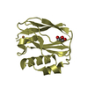
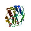
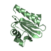
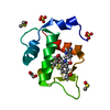
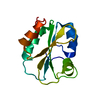
| Entry | Database: PDB / ID: 2z0t | ||||||
|---|---|---|---|---|---|---|---|
| Title | Crystal structure of hypothetical protein PH0355 | ||||||
 Components Components | Putative uncharacterized protein PH0355 | ||||||
 Keywords Keywords |  RNA BINDING PROTEIN / alpha/beta protein / Structural Genomics / NPPSFA / National Project on Protein Structural and Functional Analyses / RIKEN Structural Genomics/Proteomics Initiative / RSGI RNA BINDING PROTEIN / alpha/beta protein / Structural Genomics / NPPSFA / National Project on Protein Structural and Functional Analyses / RIKEN Structural Genomics/Proteomics Initiative / RSGI | ||||||
| Function / homology |  Function and homology information Function and homology informationUncharacterised conserved protein UCP016134 / Hypothetical protein. / ASCH / ASCH domain / ASCH domain / Archaeosine Trna-guanine Transglycosylase; Chain: A, domain 4 / PUA-like superfamily / Roll / Mainly BetaSimilarity search - Domain/homology | ||||||
| Biological species |   Pyrococcus horikoshii (archaea) Pyrococcus horikoshii (archaea) | ||||||
| Method | X-RAY DIFFRACTION / SYNCHROTRON / MAD / Resolution: 1.8 Å | ||||||
 Authors Authors | Murayama, K. / Takemoto, C. / Kato-Murayama, M. / Kawazoe, M. / Terada, T. / Shirouzu, M. / Yokoyama, S. / RIKEN Structural Genomics/Proteomics Initiative (RSGI) | ||||||
 Citation Citation | Journal: To be Published Title: Crystal structure of hypothetical protein PH0355 Authors: Murayama, K. / Takemoto, C. / Kato-Murayma, M. / Kawazoe, M. / Terada, T. / Shirouzu, M. / Yokoyama, S. | ||||||
| History |
|
- Structure visualization
| Structure viewer | Molecule: MolmilJmol/JSmol |
|---|
- Downloads & links
-Download
| PDBx/mmCIF format | 2z0t.cif.gz | 110 KB | Display | PDBx/mmCIF format |
|---|---|---|---|---|
| PDB format | pdb2z0t.ent.gz | 86.6 KB | Display | PDB format |
| PDBx/mmJSON format | 2z0t.json.gz | Tree view | PDBx/mmJSON format | |
| Others |  Other downloads Other downloads |
-Validation report
| Arichive directory | https://data.pdbj.org/pub/pdb/validation_reports/z0/2z0tftp://data.pdbj.org/pub/pdb/validation_reports/z0/2z0t | HTTPS FTP |
|---|
-Related structure data
| Similar structure data | |
|---|---|
| Other databases |


-Links
 PDBj
PDBj
- Assembly
| Deposited unit | 
| ||||||||
|---|---|---|---|---|---|---|---|---|---|
| 1 | 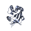
| ||||||||
| 2 | 
| ||||||||
| 3 | 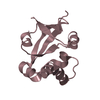
| ||||||||
| 4 | 
| ||||||||
| Unit cell |
| ||||||||
| Details | AUTHOR DETERMINED BIOLOGICAL UNIT: UNKNOWN |
-Components
| #1: Protein | Mass: 12818.990 Da / Num. of mol.: 4 Source method: isolated from a genetically manipulated source Source: (gene. exp.) Pyrococcus horikoshii (archaea) / Plasmid: pET11a / Production host:  Escherichia coli (E. coli) / References: UniProt: O58093 Escherichia coli (E. coli) / References: UniProt: O58093#2: Water | ChemComp-HOH / | Water Mass: 18.015 Da / Num. of mol.: 606 / Source method: isolated from a natural source / Formula: H2O Mass: 18.015 Da / Num. of mol.: 606 / Source method: isolated from a natural source / Formula: H2O |
|---|
-Experimental details
-Experiment
| Experiment | Method: X-RAY DIFFRACTION / Number of used crystals: 2 |
|---|
- Sample preparation
| Crystal | Density Matthews: 2.37 Å3/Da / Density % sol: 48.07 % |
|---|
-Data collection
| Diffraction |
| ||||||||||||||||||
|---|---|---|---|---|---|---|---|---|---|---|---|---|---|---|---|---|---|---|---|
| Diffraction source |
| ||||||||||||||||||
| Detector |
| ||||||||||||||||||
| Radiation |
| ||||||||||||||||||
| Radiation wavelength |
| ||||||||||||||||||
| Reflection | Resolution: 1.8→50 Å / Num. obs: 41140 / % possible obs: 94 % / Observed criterion σ(F): -3 / Redundancy: 4 % / Biso Wilson estimate: 17 Å2 / Rsym value: 0.031 / Net I/σ(I): 33.8 | ||||||||||||||||||
| Reflection shell | Resolution: 1.8→1.86 Å / Rsym value: 0.092 / % possible all: 90.2 |

- Processing
| Software |
| ||||||||||||||||||||||||||||||||||||||||||||||||||||||||||||||||||||||||||||||||
|---|---|---|---|---|---|---|---|---|---|---|---|---|---|---|---|---|---|---|---|---|---|---|---|---|---|---|---|---|---|---|---|---|---|---|---|---|---|---|---|---|---|---|---|---|---|---|---|---|---|---|---|---|---|---|---|---|---|---|---|---|---|---|---|---|---|---|---|---|---|---|---|---|---|---|---|---|---|---|---|---|---|
| Refinement | Method to determine structure: MAD / Resolution: 1.8→25.66 Å / Rfactor Rfree error: 0.005 / Data cutoff high absF: 418886.46 / Data cutoff low absF: 0 / Isotropic thermal model: RESTRAINED / Cross valid method: THROUGHOUT / σ(F): 0
| ||||||||||||||||||||||||||||||||||||||||||||||||||||||||||||||||||||||||||||||||
| Solvent computation | Solvent model: FLAT MODEL / Bsol: 41.1191 Å2 / ksol: 0.32389 e/Å3 | ||||||||||||||||||||||||||||||||||||||||||||||||||||||||||||||||||||||||||||||||
| Displacement parameters | Biso mean: 21.8 Å2
| ||||||||||||||||||||||||||||||||||||||||||||||||||||||||||||||||||||||||||||||||
| Refine analyze |
| ||||||||||||||||||||||||||||||||||||||||||||||||||||||||||||||||||||||||||||||||
| Refinement step | Cycle: LAST / Resolution: 1.8→25.66 Å
| ||||||||||||||||||||||||||||||||||||||||||||||||||||||||||||||||||||||||||||||||
| Refine LS restraints |
| ||||||||||||||||||||||||||||||||||||||||||||||||||||||||||||||||||||||||||||||||
| LS refinement shell | Resolution: 1.8→1.91 Å / Rfactor Rfree error: 0.013 / Total num. of bins used: 6
| ||||||||||||||||||||||||||||||||||||||||||||||||||||||||||||||||||||||||||||||||
| Xplor file |
|
