 Movie
Movie Controller

[English] 日本語
 Yorodumi
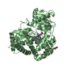
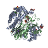
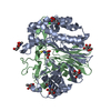
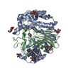
- PDB-4gdx: Crystal Structure of Human Gamma-Glutamyl Transpeptidase--Glutama... -
+ Open data

- Basic information
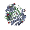
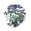
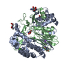
| Entry | Database: PDB / ID: 4gdx | ||||||
|---|---|---|---|---|---|---|---|
| Title | Crystal Structure of Human Gamma-Glutamyl Transpeptidase--Glutamate complex | ||||||
 Components Components | (Gamma-glutamyltranspeptidase 1 ...) x 2 | ||||||
 Keywords Keywords |  HYDROLASE / product-enzyme complex / Ntn-hydrolase family / glycoprotein / N-glycosylation / cell surface HYDROLASE / product-enzyme complex / Ntn-hydrolase family / glycoprotein / N-glycosylation / cell surface | ||||||
| Function / homology |  Function and homology informationleukotriene-C4 hydrolase / peptidyltransferase activity / leukotriene-C(4) hydrolase / leukotriene D4 biosynthetic process / Defective GGT1 causes GLUTH / Defective GGT1 in aflatoxin detoxification causes GLUTH / LTC4-CYSLTR mediated IL4 production / Glutathione synthesis and recycling / peptide modification / gamma-glutamyltransferase ...leukotriene-C4 hydrolase / peptidyltransferase activity / leukotriene-C(4) hydrolase / leukotriene D4 biosynthetic process / Defective GGT1 causes GLUTH / Defective GGT1 in aflatoxin detoxification causes GLUTH / LTC4-CYSLTR mediated IL4 production / Glutathione synthesis and recycling / peptide modification / gamma-glutamyltransferase / glutathione gamma-glutamate hydrolase / glutathione hydrolase activity / leukotriene C4 gamma-glutamyl transferase activity / glutathione catabolic process / glutathione biosynthetic process / glutamate metabolic process / leukotriene metabolic process / Aflatoxin activation and detoxification / cysteine biosynthetic process / Synthesis of Leukotrienes (LT) and Eoxins (EX) / regulation of immune system process / Paracetamol ADME / zymogen activation / amino acid metabolic process / fatty acid metabolic process / regulation of inflammatory response / spermatogenesis / proteolysis / extracellular space / extracellular exosome / plasma membrane Function and homology informationleukotriene-C4 hydrolase / peptidyltransferase activity / leukotriene-C(4) hydrolase / leukotriene D4 biosynthetic process / Defective GGT1 causes GLUTH / Defective GGT1 in aflatoxin detoxification causes GLUTH / LTC4-CYSLTR mediated IL4 production / Glutathione synthesis and recycling / peptide modification / gamma-glutamyltransferase ...leukotriene-C4 hydrolase / peptidyltransferase activity / leukotriene-C(4) hydrolase / leukotriene D4 biosynthetic process / Defective GGT1 causes GLUTH / Defective GGT1 in aflatoxin detoxification causes GLUTH / LTC4-CYSLTR mediated IL4 production / Glutathione synthesis and recycling / peptide modification / gamma-glutamyltransferase / glutathione gamma-glutamate hydrolase / glutathione hydrolase activity / leukotriene C4 gamma-glutamyl transferase activity / glutathione catabolic process / glutathione biosynthetic process / glutamate metabolic process / leukotriene metabolic process / Aflatoxin activation and detoxification / cysteine biosynthetic process / Synthesis of Leukotrienes (LT) and Eoxins (EX) / regulation of immune system process / Paracetamol ADME / zymogen activation / amino acid metabolic process / fatty acid metabolic process / regulation of inflammatory response / spermatogenesis / proteolysis / extracellular space / extracellular exosome / plasma membraneSimilarity search - Function | ||||||
| Biological species |  Homo sapiens (human) Homo sapiens (human) | ||||||
| Method | X-RAY DIFFRACTION / SYNCHROTRON / MOLECULAR REPLACEMENT / Resolution: 1.67 Å | ||||||
 Authors Authors | West, M.B. / Chen, Y. / Wickham, S. / Heroux, A. / Cahill, K. / Hanigan, M.H. / Mooers, B.H.M. | ||||||
 Citation Citation | Journal: J.Biol.Chem. / Year: 2013 Title: Novel Insights into Eukaryotic gamma-Glutamyltranspeptidase 1 from the Crystal Structure of the Glutamate-bound Human Enzyme. Authors: West, M.B. / Chen, Y. / Wickham, S. / Heroux, A. / Cahill, K. / Hanigan, M.H. / Mooers, B.H. | ||||||
| History |
|
- Structure visualization
| Structure viewer | Molecule: MolmilJmol/JSmol |
|---|
- Downloads & links
-Download
| PDBx/mmCIF format | 4gdx.cif.gz | 231.1 KB | Display | PDBx/mmCIF format |
|---|---|---|---|---|
| PDB format | pdb4gdx.ent.gz | 186.3 KB | Display | PDB format |
| PDBx/mmJSON format | 4gdx.json.gz | Tree view | PDBx/mmJSON format | |
| Others |  Other downloads Other downloads |
-Validation report
| Arichive directory | https://data.pdbj.org/pub/pdb/validation_reports/gd/4gdxftp://data.pdbj.org/pub/pdb/validation_reports/gd/4gdx | HTTPS FTP |
|---|
-Related structure data
| Related structure data |  4gg2C 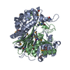 2dbuS  4gg3 S: Starting model for refinement C: citing same article ( |
|---|---|
| Similar structure data |


-Links
 PDBj
PDBj


- Assembly
| Deposited unit | 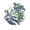
| ||||||||
|---|---|---|---|---|---|---|---|---|---|
| 1 |
| ||||||||
| Unit cell |
|
-Components
-Gamma-glutamyltranspeptidase 1 ... , 2 types, 2 molecules AB
| #1: Protein | Mass: 40874.965 Da / Num. of mol.: 1 Source method: isolated from a genetically manipulated source Source: (gene. exp.) Homo sapiens (human) / Gene: GGT, GGT1 / Production host:  PICHIA (fungus) PICHIA (fungus)References: UniProt: P19440, gamma-glutamyltransferase, glutathione gamma-glutamate hydrolase, leukotriene-C4 hydrolase |
|---|---|
| #2: Protein | Mass: 20556.938 Da / Num. of mol.: 1 Source method: isolated from a genetically manipulated source Source: (gene. exp.) Homo sapiens (human) / Gene: GGT, GGT1 / Production host: PICHIA (fungus)References: UniProt: P19440, gamma-glutamyltransferase, glutathione gamma-glutamate hydrolase, leukotriene-C4 hydrolase |
-Sugars , 1 types, 6 molecules 
| #3: Sugar | ChemComp-NAG / N-Acetylglucosamine Type: D-saccharide, beta linking / Mass: 221.208 Da / Num. of mol.: 6 Type: D-saccharide, beta linking / Mass: 221.208 Da / Num. of mol.: 6Source method: isolated from a genetically manipulated source Formula: C8H15NO6 |
|---|
-Non-polymers , 4 types, 603 molecules 


| #4: Chemical | Chloride Mass: 35.453 Da / Num. of mol.: 2 / Source method: obtained synthetically / Formula: Cl Mass: 35.453 Da / Num. of mol.: 2 / Source method: obtained synthetically / Formula: Cl#5: Chemical | ChemComp-NA / |  Mass: 22.990 Da / Num. of mol.: 1 / Source method: obtained synthetically / Formula: Na Mass: 22.990 Da / Num. of mol.: 1 / Source method: obtained synthetically / Formula: Na#6: Chemical | ChemComp-GLU / | Glutamic acid Type: L-peptide linking / Mass: 147.129 Da / Num. of mol.: 1 / Source method: obtained synthetically / Formula: C5H9NO4 Type: L-peptide linking / Mass: 147.129 Da / Num. of mol.: 1 / Source method: obtained synthetically / Formula: C5H9NO4#7: Water | ChemComp-HOH / | WaterMass: 18.015 Da / Num. of mol.: 599 / Source method: isolated from a natural source / Formula: H2O |
|---|
-Details
| Sequence details | THE UNP P19440 HAS VAL272 WHEN THIS STRUCTURE HAS ALA272. ALA272 IS A NATURAL VARIANT ON THIS HUMAN ...THE UNP P19440 HAS VAL272 WHEN THIS STRUCTURE HAS ALA272. ALA272 IS A NATURAL VARIANT ON THIS HUMAN PROTEIN. THE SEQUENCE OF THIS VARIANT IS GIVEN BY GENBANK AAA52546.1 |
|---|
-Experimental details
-Experiment
| Experiment | Method: X-RAY DIFFRACTION / Number of used crystals: 1 |
|---|
- Sample preparation
| Crystal | Density Matthews: 2.81 Å3/Da / Density % sol: 56.22 % |
|---|---|
| Crystal grow | Temperature: 298 K / Method: vapor diffusion, hanging drop / pH: 7 Details: 17.5% PEG3350, 100 mM Ammonium chloride, 0.5 mM L-glutamate, and 100 mM Na:Cacodylate pH 6.0 , VAPOR DIFFUSION, HANGING DROP, temperature 298K |
-Data collection
| Diffraction | Mean temperature: 100 K |
|---|---|
| Diffraction source | Source: SYNCHROTRON / Site: NSLS  / Beamline: X25 / Wavelength: 1.1 Å / Beamline: X25 / Wavelength: 1.1 Å |
| Detector | Type: DECTRIS PILATUS 6M / Detector: PIXEL / Date: Oct 18, 2011 Details: A Si-111 double crystal monochromator that provides a broad range 7-15 keV (0.8 2-1.8 ) x-rays. The undulator beam is focused vertically by a Rh-coated mirror. |
| Radiation | Monochromator: A Si-111 double crystal monochromator that provides a broad range 7-15 keV (0.82-1.8 ) x-rays. The undulator beam is focused vertically by a Rh-coated mirror. Protocol: SINGLE WAVELENGTH / Monochromatic (M) / Laue (L): M / Scattering type: x-ray |
| Radiation wavelength | Wavelength: 1.1 Å / Relative weight: 1 |
| Reflection | Resolution: 1.67→47.07 Å / Num. all: 70751 / Num. obs: 70751 / % possible obs: 98.3 % / Observed criterion σ(F): 2 / Observed criterion σ(I): 2 / Redundancy: 6.3 % / Biso Wilson estimate: 17.2 Å2 / Rmerge(I) obs: 0.076 / Rsym value: 0.076 / Net I/σ(I): 16.2 |
| Reflection shell | Resolution: 1.67→1.76 Å / Redundancy: 4.6 % / Rmerge(I) obs: 0.74 / Mean I/σ(I) obs: 2.1 / Rsym value: 0.74 / % possible all: 90.2 |
- Processing
| Software |
| |||||||||||||||||||||||||||||||||||||||||||||||||||||||||||||||||||||||||||||||||||||||||||||||||||||||||||||||||||||||||||||||||||||||||||||||||||||||||||||||||||||||||||||||||||||||||||||||||||||||||||
|---|---|---|---|---|---|---|---|---|---|---|---|---|---|---|---|---|---|---|---|---|---|---|---|---|---|---|---|---|---|---|---|---|---|---|---|---|---|---|---|---|---|---|---|---|---|---|---|---|---|---|---|---|---|---|---|---|---|---|---|---|---|---|---|---|---|---|---|---|---|---|---|---|---|---|---|---|---|---|---|---|---|---|---|---|---|---|---|---|---|---|---|---|---|---|---|---|---|---|---|---|---|---|---|---|---|---|---|---|---|---|---|---|---|---|---|---|---|---|---|---|---|---|---|---|---|---|---|---|---|---|---|---|---|---|---|---|---|---|---|---|---|---|---|---|---|---|---|---|---|---|---|---|---|---|---|---|---|---|---|---|---|---|---|---|---|---|---|---|---|---|---|---|---|---|---|---|---|---|---|---|---|---|---|---|---|---|---|---|---|---|---|---|---|---|---|---|---|---|---|---|---|---|---|---|
| Refinement | Method to determine structure: MOLECULAR REPLACEMENT Starting model: pdb entry 2DBU Resolution: 1.67→47.07 Å / SU ML: 0.15 / σ(F): 1.34 / Phase error: 17.18 / Stereochemistry target values: ML
| |||||||||||||||||||||||||||||||||||||||||||||||||||||||||||||||||||||||||||||||||||||||||||||||||||||||||||||||||||||||||||||||||||||||||||||||||||||||||||||||||||||||||||||||||||||||||||||||||||||||||||
| Solvent computation | Shrinkage radii: 0.9 Å / VDW probe radii: 1.11 Å / Solvent model: FLAT BULK SOLVENT MODEL | |||||||||||||||||||||||||||||||||||||||||||||||||||||||||||||||||||||||||||||||||||||||||||||||||||||||||||||||||||||||||||||||||||||||||||||||||||||||||||||||||||||||||||||||||||||||||||||||||||||||||||
| Refinement step | Cycle: LAST / Resolution: 1.67→47.07 Å
| |||||||||||||||||||||||||||||||||||||||||||||||||||||||||||||||||||||||||||||||||||||||||||||||||||||||||||||||||||||||||||||||||||||||||||||||||||||||||||||||||||||||||||||||||||||||||||||||||||||||||||
| Refine LS restraints |
| |||||||||||||||||||||||||||||||||||||||||||||||||||||||||||||||||||||||||||||||||||||||||||||||||||||||||||||||||||||||||||||||||||||||||||||||||||||||||||||||||||||||||||||||||||||||||||||||||||||||||||
| LS refinement shell |
|
