 Movie
Movie Controller

[English] 日本語
 Yorodumi
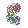
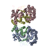
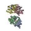
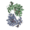
- PDB-2nqo: Crystal Structure of Helicobacter pylori gamma-Glutamyltranspeptidase -
+ Open data

- Basic information
| Entry | Database: PDB / ID: 2nqo | ||||||
|---|---|---|---|---|---|---|---|
| Title | Crystal Structure of Helicobacter pylori gamma-Glutamyltranspeptidase | ||||||
 Components Components | (Gamma-glutamyltranspeptidase) x 2 | ||||||
 Keywords Keywords |  TRANSFERASE / Ntn-hydrolase / glutamyltranspeptidase TRANSFERASE / Ntn-hydrolase / glutamyltranspeptidase | ||||||
| Function / homology |  Function and homology informationgamma-glutamyltransferase / glutathione gamma-glutamate hydrolase / glutathione hydrolase activity / leukotriene C4 gamma-glutamyl transferase activity / glutathione catabolic process / negative regulation of cell cycle G1/S phase transition / glutathione biosynthetic process / negative regulation of T cell proliferation / positive regulation of interleukin-8 production Function and homology informationgamma-glutamyltransferase / glutathione gamma-glutamate hydrolase / glutathione hydrolase activity / leukotriene C4 gamma-glutamyl transferase activity / glutathione catabolic process / negative regulation of cell cycle G1/S phase transition / glutathione biosynthetic process / negative regulation of T cell proliferation / positive regulation of interleukin-8 productionSimilarity search - Function | ||||||
| Biological species |   Helicobacter pylori (bacteria) Helicobacter pylori (bacteria) | ||||||
| Method | X-RAY DIFFRACTION / SYNCHROTRON / MOLECULAR REPLACEMENT / Resolution: 1.9 Å | ||||||
 Authors Authors | Boanca, G. / Sand, A. / Okada, T. / Suzuki, H. / Kumagai, H. / Fukuyama, K. / Barycki, J.J. | ||||||
 Citation Citation | Journal: J.Biol.Chem. / Year: 2007 Title: Autoprocessing of Helicobacter pylori gamma-glutamyltranspeptidase leads to the formation of a threonine-threonine catalytic dyad. Authors: Boanca, G. / Sand, A. / Okada, T. / Suzuki, H. / Kumagai, H. / Fukuyama, K. / Barycki, J.J. | ||||||
| History |
| ||||||
| Remark 400 | COMPOUND THE ASYMMETRIC UNIT CONTAINS A HETEROTETRAMER WHICH IS FORMED FROM TWO COMPLETE ... COMPOUND THE ASYMMETRIC UNIT CONTAINS A HETEROTETRAMER WHICH IS FORMED FROM TWO COMPLETE POLYPEPTIDE CHAINS THAT UNDERGO AUTOPROCESSING (CHAIN BREAKING BETWEEN RESIDUES 379 AND 380). THIS CHAIN BREAKING LEADS TO ACTIVATION OF THE ENZYME. AUTHOR STATES, THAT EXCEPT BEING A TRANSFERASE, THIS ENZYME IS ALSO A HYDROLASE. |
- Structure visualization
| Structure viewer | Molecule: MolmilJmol/JSmol |
|---|
- Downloads & links
-Download
| PDBx/mmCIF format | 2nqo.cif.gz | 222.5 KB | Display | PDBx/mmCIF format |
|---|---|---|---|---|
| PDB format | pdb2nqo.ent.gz | 176.8 KB | Display | PDB format |
| PDBx/mmJSON format | 2nqo.json.gz | Tree view | PDBx/mmJSON format | |
| Others |  Other downloads Other downloads |
-Validation report
| Arichive directory | https://data.pdbj.org/pub/pdb/validation_reports/nq/2nqoftp://data.pdbj.org/pub/pdb/validation_reports/nq/2nqo | HTTPS FTP |
|---|
-Related structure data
| Related structure data | 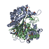 2dbuS S: Starting model for refinement |
|---|---|
| Similar structure data |


-Links
 PDBj
PDBj


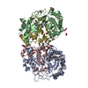
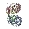
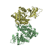
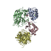
- Assembly
| Deposited unit | 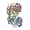
| ||||||||
|---|---|---|---|---|---|---|---|---|---|
| 1 |
| ||||||||
| 2 | 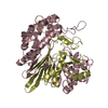
| ||||||||
| 3 | 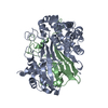
| ||||||||
| Unit cell |
|
-Components
| #1: Protein | Mass: 40506.305 Da / Num. of mol.: 2 / Fragment: Residues 27-379 / Mutation: Engineered N-terminal histidine tag Source method: isolated from a genetically manipulated source Source: (gene. exp.) Helicobacter pylori (bacteria) / Gene: HP_1118 / Plasmid: pET28a / Production host: Escherichia coli (E. coli) / Strain (production host): Rosetta2 / References: UniProt: O25743, gamma-glutamyltransferase#2: Protein | Mass: 20410.186 Da / Num. of mol.: 2 / Fragment: Residues 380-567 Source method: isolated from a genetically manipulated source Source: (gene. exp.) Helicobacter pylori (bacteria) / Gene: HP_1118 / Plasmid: pET28a / Production host: Escherichia coli (E. coli) / Strain (production host): Rosetta2 / References: UniProt: O25743, gamma-glutamyltransferase#3: Water | ChemComp-HOH / | Water Mass: 18.015 Da / Num. of mol.: 590 / Source method: isolated from a natural source / Formula: H2O Mass: 18.015 Da / Num. of mol.: 590 / Source method: isolated from a natural source / Formula: H2O |
|---|
-Experimental details
-Experiment
| Experiment | Method: X-RAY DIFFRACTION / Number of used crystals: 1 |
|---|
- Sample preparation
| Crystal | Density Matthews: 2.13 Å3/Da / Density % sol: 42.38 % |
|---|---|
| Crystal grow | Temperature: 291 K / Method: vapor diffusion, sitting drop / pH: 7.5 Details: 200 mM HEPES, 25% PEG MME2000, 5 mg/mL protein, pH 7.5, VAPOR DIFFUSION, SITTING DROP, temperature 291K |
-Data collection
| Diffraction | Mean temperature: 100 K |
|---|---|
| Diffraction source | Source: SYNCHROTRON / Site: APS  / Beamline: 14-BM-C / Wavelength: 0.9 Å / Beamline: 14-BM-C / Wavelength: 0.9 Å |
| Detector | Type: ADSC QUANTUM 315 / Detector: CCD / Date: Mar 4, 2006 |
| Radiation | Monochromator: Bent Ge(111) / Protocol: SINGLE WAVELENGTH / Monochromatic (M) / Laue (L): M / Scattering type: x-ray |
| Radiation wavelength | Wavelength: 0.9 Å / Relative weight: 1 |
| Reflection | Resolution: 1.9→100 Å / Num. all: 72974 / Num. obs: 72974 / % possible obs: 91.5 % / Observed criterion σ(F): 0 / Observed criterion σ(I): 0 / Redundancy: 4.8 % / Biso Wilson estimate: 16.2 Å2 / Rmerge(I) obs: 0.046 / Χ2: 1.035 / Net I/σ(I): 32.3 |
| Reflection shell | Resolution: 1.9→2.02 Å / Redundancy: 4.8 % / Rmerge(I) obs: 0.466 / Num. unique all: 7605 / Χ2: 1.211 / % possible all: 77.6 |
-Phasing
| Phasing MR | Method rotation: fast direct / Method translation: &STRIP%trans_method |
|---|
- Processing
| Software |
| ||||||||||||||||||||||||||||||||||||
|---|---|---|---|---|---|---|---|---|---|---|---|---|---|---|---|---|---|---|---|---|---|---|---|---|---|---|---|---|---|---|---|---|---|---|---|---|---|
| Refinement | Method to determine structure: MOLECULAR REPLACEMENT Starting model: PDB entry 2DBU Resolution: 1.9→28.19 Å / Rfactor Rfree error: 0.003 / Data cutoff high absF: 163846.344 / Data cutoff low absF: 0 / Isotropic thermal model: RESTRAINED / Cross valid method: THROUGHOUT / σ(F): 0
| ||||||||||||||||||||||||||||||||||||
| Solvent computation | Solvent model: FLAT MODEL / Bsol: 53.788 Å2 / ksol: 0.367 e/Å3 | ||||||||||||||||||||||||||||||||||||
| Displacement parameters | Biso mean: 31.9 Å2
| ||||||||||||||||||||||||||||||||||||
| Refine analyze |
| ||||||||||||||||||||||||||||||||||||
| Refinement step | Cycle: LAST / Resolution: 1.9→28.19 Å
| ||||||||||||||||||||||||||||||||||||
| Refine LS restraints |
| ||||||||||||||||||||||||||||||||||||
| LS refinement shell | Resolution: 1.9→2.02 Å / Rfactor Rfree error: 0.009 / Total num. of bins used: 6
| ||||||||||||||||||||||||||||||||||||
| Xplor file |
|
