 Movie
Movie Controller


+ Open data

- Basic information
| Entry | Database: PDB / ID: 4dum | ||||||
|---|---|---|---|---|---|---|---|
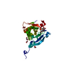
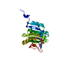
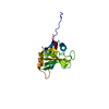
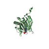
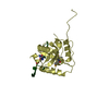
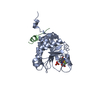
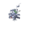
| Title | Co-crystal structure of eIF4E with inhibitor | ||||||
 Components Components | Eukaryotic translation initiation factor 4E EIF4E EIF4E | ||||||
 Keywords Keywords | TRANSLATION / CAP-binding protein / translation initiation factor / m7GTP | ||||||
| Function / homology |  Function and homology information Function and homology informationActivation of the mRNA upon binding of the cap-binding complex and eIFs, and subsequent binding to 43S / : / eukaryotic initiation factor 4G binding / regulation of translation at postsynapse, modulating synaptic transmission / RNA cap binding / chromatoid body / eukaryotic translation initiation factor 4F complex / Z-decay: degradation of maternal mRNAs by zygotically expressed factors / mRNA cap binding / Deadenylation of mRNA ...Activation of the mRNA upon binding of the cap-binding complex and eIFs, and subsequent binding to 43S / : / eukaryotic initiation factor 4G binding / regulation of translation at postsynapse, modulating synaptic transmission / RNA cap binding / chromatoid body / eukaryotic translation initiation factor 4F complex / Z-decay: degradation of maternal mRNAs by zygotically expressed factors / mRNA cap binding / Deadenylation of mRNA / Transport of the SLBP independent Mature mRNA / RNA 7-methylguanosine cap binding / Transport of the SLBP Dependant Mature mRNA / M-decay: degradation of maternal mRNAs by maternally stored factors / Transport of Mature mRNA Derived from an Intronless Transcript / RISC complex / Ribosomal scanning and start codon recognition / Translation initiation complex formation / stem cell population maintenance / mTORC1-mediated signalling / GTP hydrolysis and joining of the 60S ribosomal subunit / negative regulation of neuron differentiation / L13a-mediated translational silencing of Ceruloplasmin expression / behavioral fear response / mRNA export from nucleus / translational initiation / translation initiation factor activity / positive regulation of mitotic cell cycle / cellular response to dexamethasone stimulus / P-body / neuron differentiation / G1/S transition of mitotic cell cycle / ISG15 antiviral mechanism / cytoplasmic stress granule / cytoplasmic ribonucleoprotein granule / regulation of translation / postsynapse / DNA-binding transcription factor binding / negative regulation of translation / nuclear speck / glutamatergic synapse / perinuclear region of cytoplasm / enzyme binding / RNA binding / extracellular exosome / nucleus / cytosol / cytoplasmSimilarity search - Function | ||||||
| Biological species |  Homo sapiens (human) Homo sapiens (human) | ||||||
| Method | X-RAY DIFFRACTION / MOLECULAR REPLACEMENT / Resolution: 2.95 Å | ||||||
 Authors Authors | Min, X. / Johnstone, S. / Walker, N. / Wang, Z. | ||||||
 Citation Citation | Journal: J.Med.Chem. / Year: 2012 Title: Structure-Guided Design, Synthesis, and Evaluation of Guanine-Derived Inhibitors of the eIF4E mRNA-Cap Interaction. Authors: Chen, X. / Kopecky, D.J. / Mihalic, J. / Jeffries, S. / Min, X. / Heath, J. / Deignan, J. / Lai, S. / Fu, Z. / Guimaraes, C. / Shen, S. / Li, S. / Johnstone, S. / Thibault, S. / Xu, H. / ...Authors: Chen, X. / Kopecky, D.J. / Mihalic, J. / Jeffries, S. / Min, X. / Heath, J. / Deignan, J. / Lai, S. / Fu, Z. / Guimaraes, C. / Shen, S. / Li, S. / Johnstone, S. / Thibault, S. / Xu, H. / Cardozo, M. / Shen, W. / Walker, N. / Kayser, F. / Wang, Z. | ||||||
| History |
|
- Structure visualization
| Structure viewer | Molecule: MolmilJmol/JSmol |
|---|
- Downloads & links
-Download
| PDBx/mmCIF format | 4dum.cif.gz | 54.5 KB | Display | PDBx/mmCIF format |
|---|---|---|---|---|
| PDB format | pdb4dum.ent.gz | 38.6 KB | Display | PDB format |
| PDBx/mmJSON format | 4dum.json.gz | Tree view | PDBx/mmJSON format | |
| Others |  Other downloads Other downloads |
-Validation report
| Arichive directory | https://data.pdbj.org/pub/pdb/validation_reports/du/4dumftp://data.pdbj.org/pub/pdb/validation_reports/du/4dum | HTTPS FTP |
|---|
-Related structure data


-Links
 PDBj
PDBj


- Assembly
| Deposited unit | 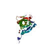
| ||||||||
|---|---|---|---|---|---|---|---|---|---|
| 1 |
| ||||||||
| Unit cell |
|
-Components
| #1: Protein | EIF4E / eIF-4E / eIF4E / eIF-4F 25 kDa subunit / mRNA cap-binding protein Mass: 28040.301 Da / Num. of mol.: 1 Source method: isolated from a genetically manipulated source Source: (gene. exp.) Homo sapiens (human) / Gene: EIF4E, EIF4EL1, EIF4F / Plasmid: pET101 / Production host:  Escherichia coli (E. coli) / References: UniProt: P06730 Escherichia coli (E. coli) / References: UniProt: P06730 | ||
|---|---|---|---|
| #2: Chemical | ChemComp-HLI / (  Mass: 475.822 Da / Num. of mol.: 1 / Source method: obtained synthetically / Formula: C20H19ClN5O5P Mass: 475.822 Da / Num. of mol.: 1 / Source method: obtained synthetically / Formula: C20H19ClN5O5P | ||
| #3: Chemical | Ethylene glycol  Mass: 62.068 Da / Num. of mol.: 2 / Source method: obtained synthetically / Formula: C2H6O2 Mass: 62.068 Da / Num. of mol.: 2 / Source method: obtained synthetically / Formula: C2H6O2#4: Water | ChemComp-HOH / | Water Mass: 18.015 Da / Num. of mol.: 14 / Source method: isolated from a natural source / Formula: H2O Mass: 18.015 Da / Num. of mol.: 14 / Source method: isolated from a natural source / Formula: H2O |
-Experimental details
-Experiment
| Experiment | Method: X-RAY DIFFRACTION / Number of used crystals: 1 |
|---|
- Sample preparation
| Crystal | Density Matthews: 2.51 Å3/Da / Density % sol: 51.04 % |
|---|---|
| Crystal grow | Temperature: 289 K / Method: vapor diffusion, sitting drop Details: The purified protein which contained 100 uM m7-GTP was then concentrated to about 7 mg/mL in 20 mM Hepes, pH7.6, 100 mM KCl, 1mM DTT, 0.1 mM EDTA for crystallization. The m7-GTP-bound eIF4e ...Details: The purified protein which contained 100 uM m7-GTP was then concentrated to about 7 mg/mL in 20 mM Hepes, pH7.6, 100 mM KCl, 1mM DTT, 0.1 mM EDTA for crystallization. The m7-GTP-bound eIF4e protein was crystallized with 1:1 ratio of protein solution to reservoir solution of 17-20% PEG-3350 and 0.1-0.4M Na formate, VAPOR DIFFUSION, SITTING DROP, temperature 289K |
-Data collection
| Diffraction | Mean temperature: 90 K |
|---|---|
| Diffraction source | Source: ROTATING ANODE / Type: RIGAKU FR-E SUPERBRIGHT / Wavelength: 1.54 Å |
| Detector | Type: RIGAKU RAXIS HTC / Detector: IMAGE PLATE / Date: May 1, 2007 / Details: Rigaku Varimax HR optics |
| Radiation | Protocol: SINGLE WAVELENGTH / Monochromatic (M) / Laue (L): M / Scattering type: x-ray |
| Radiation wavelength | Wavelength: 1.54 Å / Relative weight: 1 |
| Reflection | Resolution: 2.95→62.75 Å / Num. all: 18074 / Num. obs: 6282 / % possible obs: 98.9 % / Observed criterion σ(I): 2.9 / Redundancy: 2.9 % / Biso Wilson estimate: 58.7 Å2 / Rmerge(I) obs: 0.116 / Net I/σ(I): 9.5 |
| Reflection shell | Resolution: 2.95→3.11 Å / Rmerge(I) obs: 0.324 / Mean I/σ(I) obs: 2.3 / Num. unique all: 765 / % possible all: 97.5 |
- Processing
| Software |
| |||||||||||||||||||||||||||||||||||||||||||||||||||||||||||||||||
|---|---|---|---|---|---|---|---|---|---|---|---|---|---|---|---|---|---|---|---|---|---|---|---|---|---|---|---|---|---|---|---|---|---|---|---|---|---|---|---|---|---|---|---|---|---|---|---|---|---|---|---|---|---|---|---|---|---|---|---|---|---|---|---|---|---|---|
| Refinement | Method to determine structure: MOLECULAR REPLACEMENT / Resolution: 2.95→38.236 Å / Cor.coef. Fo:Fc: 0.918 / Cor.coef. Fo:Fc free: 0.852 / SU B: 16.91 / SU ML: 0.317 / Cross valid method: THROUGHOUT / ESU R Free: 0.425 / Stereochemistry target values: MAXIMUM LIKELIHOOD / Details: HYDROGENS HAVE BEEN ADDED IN THE RIDING POSITIONS
| |||||||||||||||||||||||||||||||||||||||||||||||||||||||||||||||||
| Solvent computation | Ion probe radii: 0.8 Å / Shrinkage radii: 0.8 Å / VDW probe radii: 1.4 Å / Solvent model: MASK | |||||||||||||||||||||||||||||||||||||||||||||||||||||||||||||||||
| Displacement parameters | Biso mean: 35.343 Å2
| |||||||||||||||||||||||||||||||||||||||||||||||||||||||||||||||||
| Refinement step | Cycle: LAST / Resolution: 2.95→38.236 Å
| |||||||||||||||||||||||||||||||||||||||||||||||||||||||||||||||||
| Refine LS restraints |
| |||||||||||||||||||||||||||||||||||||||||||||||||||||||||||||||||
| LS refinement shell | Resolution: 2.95→3.027 Å / Total num. of bins used: 20
|
