 Movie
Movie Controller


+ Open data

- Basic information
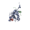
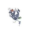
| Entry | Database: PDB / ID: 5ei3 | |||||||||
|---|---|---|---|---|---|---|---|---|---|---|
| Title | Co-crystal structure of eIF4E with nucleotide mimetic inhibitor. | |||||||||
 Components Components |
| |||||||||
 Keywords Keywords |  TRANSLATION / Complex / inhibitor / eIF4E TRANSLATION / Complex / inhibitor / eIF4E | |||||||||
| Function / homology |  Function and homology information Function and homology informationpositive regulation of eukaryotic translation initiation factor 4F complex assembly / positive regulation of mRNA cap binding / positive regulation of translation in response to endoplasmic reticulum stress / cap-dependent translational initiation / macromolecule biosynthetic process / Activation of the mRNA upon binding of the cap-binding complex and eIFs, and subsequent binding to 43S / eukaryotic initiation factor 4G binding / eukaryotic initiation factor 4E binding / regulation of cellular response to stress / regulation of translation at postsynapse, modulating synaptic transmission ...positive regulation of eukaryotic translation initiation factor 4F complex assembly / positive regulation of mRNA cap binding / positive regulation of translation in response to endoplasmic reticulum stress / cap-dependent translational initiation / macromolecule biosynthetic process / Activation of the mRNA upon binding of the cap-binding complex and eIFs, and subsequent binding to 43S / eukaryotic initiation factor 4G binding / eukaryotic initiation factor 4E binding / regulation of cellular response to stress / regulation of translation at postsynapse, modulating synaptic transmission / RNA cap binding / chromatoid body / eukaryotic translation initiation factor 4F complex / Z-decay: degradation of maternal mRNAs by zygotically expressed factors / translation factor activity, RNA binding / mRNA cap binding / : / Deadenylation of mRNA / miRNA-mediated gene silencing by inhibition of translation / RNA 7-methylguanosine cap binding / Transport of the SLBP independent Mature mRNA / Transport of the SLBP Dependant Mature mRNA / M-decay: degradation of maternal mRNAs by maternally stored factors / Transport of Mature mRNA Derived from an Intronless Transcript / positive regulation of protein localization to cell periphery / RISC complex / regulation of translational initiation / Ribosomal scanning and start codon recognition / Translation initiation complex formation / stem cell population maintenance / negative regulation of peptidyl-threonine phosphorylation / mTORC1-mediated signalling / cellular response to nutrient levels / regulation of presynapse assembly / Nonsense Mediated Decay (NMD) independent of the Exon Junction Complex (EJC) / GTP hydrolysis and joining of the 60S ribosomal subunit / negative regulation of neuron differentiation / L13a-mediated translational silencing of Ceruloplasmin expression / positive regulation of G1/S transition of mitotic cell cycle / behavioral fear response / Nonsense Mediated Decay (NMD) enhanced by the Exon Junction Complex (EJC) / mRNA export from nucleus / translation initiation factor binding / energy homeostasis / translational initiation / translation initiation factor activity / positive regulation of protein metabolic process / positive regulation of neuron differentiation / cellular response to dexamethasone stimulus / positive regulation of mitotic cell cycle / negative regulation of autophagy / AUF1 (hnRNP D0) binds and destabilizes mRNA / P-body / lung development / G1/S transition of mitotic cell cycle / cytoplasmic ribonucleoprotein granule / neuron differentiation / ISG15 antiviral mechanism / Regulation of expression of SLITs and ROBOs / cytoplasmic stress granule / regulation of translation / positive regulation of peptidyl-serine phosphorylation / postsynapse / positive regulation of cell growth / response to ethanol / DNA-binding transcription factor binding / negative regulation of translation / molecular adaptor activity / ribosome / nuclear speck / translation / mRNA binding / glutamatergic synapse / perinuclear region of cytoplasm / enzyme binding / RNA binding / extracellular exosome / ATP binding / membrane / identical protein binding / nucleus / cytosol / cytoplasmSimilarity search - Function | |||||||||
| Biological species |  Homo sapiens (human) Homo sapiens (human)synthetic construct (others) | |||||||||
| Method | X-RAY DIFFRACTION / SYNCHROTRON / MOLECULAR REPLACEMENT / Resolution: 1.71 Å | |||||||||
 Authors Authors | Nowicki, M.W. / Walkinshaw, M.D. / Fischer, P.M. | |||||||||
 Citation Citation | Journal: Eur.J.Med.Chem. / Year: 2016 Title: Design of nucleotide-mimetic and non-nucleotide inhibitors of the translation initiation factor eIF4E: Synthesis, structural and functional characterisation. Authors: Soukarieh, F. / Nowicki, M.W. / Bastide, A. / Poyry, T. / Jones, C. / Dudek, K. / Patwardhan, G. / Meullenet, F. / Oldham, N.J. / Walkinshaw, M.D. / Willis, A.E. / Fischer, P.M. | |||||||||
| History |
|
- Structure visualization
| Structure viewer | Molecule: MolmilJmol/JSmol |
|---|
- Downloads & links
-Download
| PDBx/mmCIF format | 5ei3.cif.gz | 68.3 KB | Display | PDBx/mmCIF format |
|---|---|---|---|---|
| PDB format | pdb5ei3.ent.gz | 47.7 KB | Display | PDB format |
| PDBx/mmJSON format | 5ei3.json.gz | Tree view | PDBx/mmJSON format | |
| Others |  Other downloads Other downloads |
-Validation report
| Arichive directory | https://data.pdbj.org/pub/pdb/validation_reports/ei/5ei3ftp://data.pdbj.org/pub/pdb/validation_reports/ei/5ei3 | HTTPS FTP |
|---|
-Related structure data
| Related structure data | 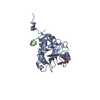 5ehcC 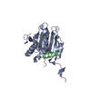 5eirC 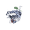 5ekvC 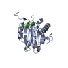 2v8wS S: Starting model for refinement C: citing same article ( |
|---|---|
| Similar structure data |


-Links
 PDBj
PDBj


- Assembly
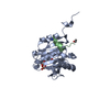
| Deposited unit | 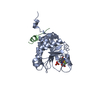
| ||||||||
|---|---|---|---|---|---|---|---|---|---|
| 1 |
| ||||||||
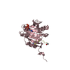
| Unit cell |
| ||||||||
| Components on special symmetry positions |
|
-Components
| #1: Protein | EIF4E / eIF4E / eIF-4F 25 kDa subunit / mRNA cap-binding protein Mass: 25130.242 Da / Num. of mol.: 1 Source method: isolated from a genetically manipulated source Source: (gene. exp.) Homo sapiens (human) / Gene: EIF4E, EIF4EL1, EIF4F / Plasmid: pET11d / Production host:  Escherichia coli BL21(DE3) (bacteria) / Variant (production host): Rosetta PLysS / References: UniProt: P06730 Escherichia coli BL21(DE3) (bacteria) / Variant (production host): Rosetta PLysS / References: UniProt: P06730 |
|---|---|
| #2: Protein/peptide | Eukaryotic translation initiation factor 4 gamma 1 Mass: 1851.155 Da / Num. of mol.: 1 / Fragment: eIF4E binding sequence / Source method: obtained synthetically / Details: commercial synthesis / Source: (synth.) synthetic construct (others) / References: UniProt: Q04637*PLUS |
| #3: Chemical | ChemComp-5O8 / ~{  Mass: 505.448 Da / Num. of mol.: 1 / Source method: obtained synthetically / Formula: C18H20F3N6O6S Mass: 505.448 Da / Num. of mol.: 1 / Source method: obtained synthetically / Formula: C18H20F3N6O6S |
| #4: Chemical | ChemComp-SO4 / Sulfate  Mass: 96.063 Da / Num. of mol.: 1 / Source method: obtained synthetically / Formula: SO4 Mass: 96.063 Da / Num. of mol.: 1 / Source method: obtained synthetically / Formula: SO4 |
| #5: Water | ChemComp-HOH / Water Mass: 18.015 Da / Num. of mol.: 355 / Source method: isolated from a natural source / Formula: H2O Mass: 18.015 Da / Num. of mol.: 355 / Source method: isolated from a natural source / Formula: H2O |
-Experimental details
-Experiment
| Experiment | Method: X-RAY DIFFRACTION / Number of used crystals: 1 |
|---|
- Sample preparation
| Crystal | Density Matthews: 2.33 Å3/Da / Density % sol: 47.24 % |
|---|---|
| Crystal grow | Temperature: 291 K / Method: vapor diffusion, hanging drop / pH: 7.5 Details: 21-31% PEG 8000, 1???3% (NH4)2SO4, 100 mM HEPES, pH 7.5, 1 round of seeding |
-Data collection
| Diffraction | Mean temperature: 100 K | ||||||||||||||||||||||||||||||||||||||||||||||||||||||||||||||||||||||||||||||||||||||||||||||||||||||||||||||
|---|---|---|---|---|---|---|---|---|---|---|---|---|---|---|---|---|---|---|---|---|---|---|---|---|---|---|---|---|---|---|---|---|---|---|---|---|---|---|---|---|---|---|---|---|---|---|---|---|---|---|---|---|---|---|---|---|---|---|---|---|---|---|---|---|---|---|---|---|---|---|---|---|---|---|---|---|---|---|---|---|---|---|---|---|---|---|---|---|---|---|---|---|---|---|---|---|---|---|---|---|---|---|---|---|---|---|---|---|---|---|---|
| Diffraction source | Source: SYNCHROTRON / Site: Diamond  / Beamline: I02 / Wavelength: 0.85 Å / Beamline: I02 / Wavelength: 0.85 Å | ||||||||||||||||||||||||||||||||||||||||||||||||||||||||||||||||||||||||||||||||||||||||||||||||||||||||||||||
| Detector | Type: ADSC QUANTUM 315 / Detector: CCD / Date: Apr 30, 2011 / Details: PSI PILATUS 6M | ||||||||||||||||||||||||||||||||||||||||||||||||||||||||||||||||||||||||||||||||||||||||||||||||||||||||||||||
| Radiation | Protocol: SINGLE WAVELENGTH / Monochromatic (M) / Laue (L): M / Scattering type: x-ray | ||||||||||||||||||||||||||||||||||||||||||||||||||||||||||||||||||||||||||||||||||||||||||||||||||||||||||||||
| Radiation wavelength | Wavelength: 0.85 Å / Relative weight: 1 | ||||||||||||||||||||||||||||||||||||||||||||||||||||||||||||||||||||||||||||||||||||||||||||||||||||||||||||||
| Reflection | Resolution: 1.71→62.595 Å / Num. all: 28165 / Num. obs: 28165 / % possible obs: 100 % / Redundancy: 7 % / Biso Wilson estimate: 11.24 Å2 / Rpim(I) all: 0.039 / Rrim(I) all: 0.102 / Rsym value: 0.094 / Net I/av σ(I): 4.843 / Net I/σ(I): 15.7 / Num. measured all: 195799 | ||||||||||||||||||||||||||||||||||||||||||||||||||||||||||||||||||||||||||||||||||||||||||||||||||||||||||||||
| Reflection shell | Diffraction-ID: 1 / Rejects: 0
|
- Processing
| Software |
| ||||||||||||||||||||||||||||||||||||||||||||||||||||||||||||||||||
|---|---|---|---|---|---|---|---|---|---|---|---|---|---|---|---|---|---|---|---|---|---|---|---|---|---|---|---|---|---|---|---|---|---|---|---|---|---|---|---|---|---|---|---|---|---|---|---|---|---|---|---|---|---|---|---|---|---|---|---|---|---|---|---|---|---|---|---|
| Refinement | Method to determine structure: MOLECULAR REPLACEMENT Starting model: 2v8w Resolution: 1.71→40.035 Å / SU ML: 0.16 / Cross valid method: NONE / σ(F): 1.7 / Phase error: 16.73 / Stereochemistry target values: ML
| ||||||||||||||||||||||||||||||||||||||||||||||||||||||||||||||||||
| Solvent computation | Shrinkage radii: 0.9 Å / VDW probe radii: 1.11 Å / Solvent model: FLAT BULK SOLVENT MODEL | ||||||||||||||||||||||||||||||||||||||||||||||||||||||||||||||||||
| Displacement parameters | Biso max: 72.63 Å2 / Biso mean: 16.2 Å2 / Biso min: 3.48 Å2 | ||||||||||||||||||||||||||||||||||||||||||||||||||||||||||||||||||
| Refinement step | Cycle: final / Resolution: 1.71→40.035 Å
| ||||||||||||||||||||||||||||||||||||||||||||||||||||||||||||||||||
| Refine LS restraints |
| ||||||||||||||||||||||||||||||||||||||||||||||||||||||||||||||||||
| LS refinement shell | Refine-ID: X-RAY DIFFRACTION / Total num. of bins used: 10 / % reflection obs: 100 %
|
