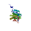
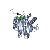
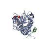
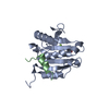
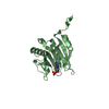
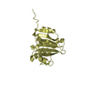
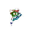
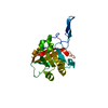
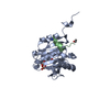
Entry Database : PDB / ID : 4azaTitle Improved eIF4E binding peptides by phage display guided design. EIF4G1_D5S PEPTIDE EUKARYOTIC TRANSLATION INITIATION FACTOR 4E Keywords Function / homology Function Domain/homology Component
/ / / / / / / / / / / / / / / / / / / / / / / / / / / / / / / / / / / / / / / / / / / / / / / / / / / / / / / / / / / / / / / / / / / / / / / / / / / / / / / / / / / / / / / / / / / / / / / / / / / / / / / / / / / / / / / / / / / / / / / / / Biological species HOMO SAPIENS (human)Method / / Resolution : 2.16 Å Authors Chew, W.Z. / Quah, S.T. / Verma, C.S. / Liu, Y. / Lane, D.P. / Brown, C.J. Journal : Plos One / Year : 2012Title : Improved Eif4E Binding Peptides by Phage Display Guided Design: Plasticity of Interacting Surfaces Yield Collective Effects.Authors : Zhou, W. / Quah, S.T. / Verma, C.S. / Liu, Y. / Lane, D.P. / Brown, C.J. History Deposition Jun 25, 2012 Deposition site / Processing site Revision 1.0 Aug 8, 2012 Provider / Type Revision 1.1 Mar 6, 2013 Group Revision 1.2 Dec 20, 2023 Group Data collection / Database references ... Data collection / Database references / Derived calculations / Other / Refinement description Category chem_comp_atom / chem_comp_bond ... chem_comp_atom / chem_comp_bond / database_2 / pdbx_database_status / pdbx_initial_refinement_model / struct_conn / struct_site Item _database_2.pdbx_DOI / _database_2.pdbx_database_accession ... _database_2.pdbx_DOI / _database_2.pdbx_database_accession / _pdbx_database_status.status_code_sf / _struct_conn.pdbx_leaving_atom_flag / _struct_conn.ptnr1_auth_comp_id / _struct_conn.ptnr1_auth_seq_id / _struct_conn.ptnr1_label_atom_id / _struct_conn.ptnr1_label_comp_id / _struct_conn.ptnr1_label_seq_id / _struct_conn.ptnr2_auth_comp_id / _struct_conn.ptnr2_auth_seq_id / _struct_conn.ptnr2_label_atom_id / _struct_conn.ptnr2_label_comp_id / _struct_conn.ptnr2_label_seq_id / _struct_site.pdbx_auth_asym_id / _struct_site.pdbx_auth_comp_id / _struct_site.pdbx_auth_seq_id
Show all Show less
 Movie
Movie Controller


 Open data
 Basic information
 Components
 Keywords
 TRANSLATION
 Function and homology information
 Authors
 Citation
 Structure visualization
 Downloads & links
 Other downloads


 PDBj
PDBj


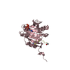
 Assembly


 Mass: 537.227 Da / Num. of mol.: 2 / Source method: obtained synthetically / Formula: C12H20N4O14P3
 Mass: 18.015 Da / Num. of mol.: 218 / Source method: isolated from a natural source / Formula: H2O
 Sample preparation
 Processing