 Movie
Movie Controller


+ Open data

- Basic information
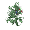
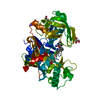
| Entry | Database: PDB / ID: 3zmg | ||||||
|---|---|---|---|---|---|---|---|
| Title | CRYSTAL STRUCTURE OF BACE-1 IN COMPLEX WITH CHEMICAL LIGAND | ||||||
 Components Components | BETA-SECRETASE 1 | ||||||
 Keywords Keywords | HYDROLASE | ||||||
| Function / homology |  Function and homology informationmemapsin 2 / Golgi-associated vesicle lumen / signaling receptor ligand precursor processing / beta-aspartyl-peptidase activity / amyloid precursor protein catabolic process / amyloid-beta formation / membrane protein ectodomain proteolysis / cellular response to manganese ion / prepulse inhibition / detection of mechanical stimulus involved in sensory perception of pain ...memapsin 2 / Golgi-associated vesicle lumen / signaling receptor ligand precursor processing / beta-aspartyl-peptidase activity / amyloid precursor protein catabolic process / amyloid-beta formation / membrane protein ectodomain proteolysis / cellular response to manganese ion / prepulse inhibition / detection of mechanical stimulus involved in sensory perception of pain / amyloid-beta metabolic process / cellular response to copper ion / hippocampal mossy fiber to CA3 synapse / presynaptic modulation of chemical synaptic transmission / multivesicular body / response to lead ion / trans-Golgi network / protein processing / recycling endosome / cellular response to amyloid-beta / positive regulation of neuron apoptotic process / synaptic vesicle / late endosome / amyloid-beta binding / peptidase activity / endopeptidase activity / amyloid fibril formation / lysosome / aspartic-type endopeptidase activity / early endosome / endosome membrane / endosome / membrane raft / Amyloid fiber formation / axon / endoplasmic reticulum lumen / neuronal cell body / dendrite / Golgi apparatus / enzyme binding / cell surface / proteolysis / membrane / plasma membrane Function and homology informationmemapsin 2 / Golgi-associated vesicle lumen / signaling receptor ligand precursor processing / beta-aspartyl-peptidase activity / amyloid precursor protein catabolic process / amyloid-beta formation / membrane protein ectodomain proteolysis / cellular response to manganese ion / prepulse inhibition / detection of mechanical stimulus involved in sensory perception of pain ...memapsin 2 / Golgi-associated vesicle lumen / signaling receptor ligand precursor processing / beta-aspartyl-peptidase activity / amyloid precursor protein catabolic process / amyloid-beta formation / membrane protein ectodomain proteolysis / cellular response to manganese ion / prepulse inhibition / detection of mechanical stimulus involved in sensory perception of pain / amyloid-beta metabolic process / cellular response to copper ion / hippocampal mossy fiber to CA3 synapse / presynaptic modulation of chemical synaptic transmission / multivesicular body / response to lead ion / trans-Golgi network / protein processing / recycling endosome / cellular response to amyloid-beta / positive regulation of neuron apoptotic process / synaptic vesicle / late endosome / amyloid-beta binding / peptidase activity / endopeptidase activity / amyloid fibril formation / lysosome / aspartic-type endopeptidase activity / early endosome / endosome membrane / endosome / membrane raft / Amyloid fiber formation / axon / endoplasmic reticulum lumen / neuronal cell body / dendrite / Golgi apparatus / enzyme binding / cell surface / proteolysis / membrane / plasma membraneSimilarity search - Function | ||||||
| Biological species |  HOMO SAPIENS (human) HOMO SAPIENS (human) | ||||||
| Method | X-RAY DIFFRACTION / SYNCHROTRON / MOLECULAR REPLACEMENT / Resolution: 1.74 Å | ||||||
 Authors Authors | Banner, D.W. / Kuglstatter, A. / Benz, J. / Stihle, M. | ||||||
 Citation Citation | Journal: J.Med.Chem. / Year: 2013 Title: Beta-Secretase (Bace1) Inhibitors with High in Vivo Efficacy Suitable for Clinical Evaluation in Alzheimer'S Disease. Authors: Hilpert, H. / Guba, W. / Woltering, T.J. / Wostl, W. / Pinard, E. / Mauser, H. / Mayweg, A.V. / Rogers-Evans, M. / Humm, R. / Krummenacher, D. / Muser, T. / Schnider, C. / Jacobsen, H. / ...Authors: Hilpert, H. / Guba, W. / Woltering, T.J. / Wostl, W. / Pinard, E. / Mauser, H. / Mayweg, A.V. / Rogers-Evans, M. / Humm, R. / Krummenacher, D. / Muser, T. / Schnider, C. / Jacobsen, H. / Ozmen, L. / Bergadano, A. / Banner, D.W. / Hochstrasser, R. / Kuglstatter, A. / David-Pierson, P. / Fischer, H. / Polara, A. / Narquizian, R. | ||||||
| History |
| ||||||
| Remark 700 | SHEET THE SHEET STRUCTURE OF THIS MOLECULE IS BIFURCATED. IN ORDER TO REPRESENT THIS FEATURE IN ... SHEET THE SHEET STRUCTURE OF THIS MOLECULE IS BIFURCATED. IN ORDER TO REPRESENT THIS FEATURE IN THE SHEET RECORDS BELOW, TWO SHEETS ARE DEFINED. |
- Structure visualization
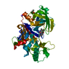
| Structure viewer | Molecule: MolmilJmol/JSmol |
|---|
- Downloads & links
-Download
| PDBx/mmCIF format | 3zmg.cif.gz | 105.1 KB | Display | PDBx/mmCIF format |
|---|---|---|---|---|
| PDB format | pdb3zmg.ent.gz | 79.8 KB | Display | PDB format |
| PDBx/mmJSON format | 3zmg.json.gz | Tree view | PDBx/mmJSON format | |
| Others |  Other downloads Other downloads |
-Validation report
| Arichive directory | https://data.pdbj.org/pub/pdb/validation_reports/zm/3zmgftp://data.pdbj.org/pub/pdb/validation_reports/zm/3zmg | HTTPS FTP |
|---|
-Related structure data
| Related structure data |  3zlqC  4j0pC  4j0tC  4j0vC  4j0yC  4j0zC  4j17C  4j1cC 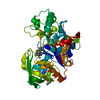 4j1eC 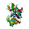 4j1fC 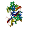 4j1hC  4j1iC 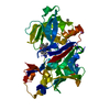 4j1kC C: citing same article ( |
|---|---|
| Similar structure data |


-Links
 PDBj
PDBj

- Assembly
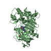
| Deposited unit | 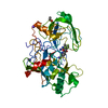
| |||||||||
|---|---|---|---|---|---|---|---|---|---|---|
| 1 |
| |||||||||
| Unit cell |
| |||||||||
| Components on special symmetry positions |
|
-Components
| #1: Protein | / 3.4.23.46 / ASPARTYL PROTEASE 2 / ASP2 / ASP 2 / BETA-SITE AMYLOID PRECURSOR PROTEIN CLEAVING ...3.4.23.46 / ASPARTYL PROTEASE 2 / ASP2 / ASP 2 / BETA-SITE AMYLOID PRECURSOR PROTEIN CLEAVING ENZYME 1 / BETA-SITE APP CLEAVING ENZYME 1 / MEMAPSIN-2 / MEMBRANE-ASSOCIATED ASPARTIC PROTEASE 2 Mass: 45554.008 Da / Num. of mol.: 1 / Fragment: EXTRACELLULAR, RESIDUES 46-454 / Mutation: YES Source method: isolated from a genetically manipulated source Source: (gene. exp.) HOMO SAPIENS (human) / Production host:  ESCHERICHIA COLI (E. coli) / References: UniProt: P56817, memapsin 2 ESCHERICHIA COLI (E. coli) / References: UniProt: P56817, memapsin 2 | ||||
|---|---|---|---|---|---|
| #2: Chemical | ChemComp-6Z0 /   Mass: 389.331 Da / Num. of mol.: 1 / Source method: obtained synthetically / Formula: C18H14F3N5O2 Mass: 389.331 Da / Num. of mol.: 1 / Source method: obtained synthetically / Formula: C18H14F3N5O2 | ||||
| #3: Chemical |   Mass: 22.990 Da / Num. of mol.: 2 / Source method: obtained synthetically / Formula: Na Mass: 22.990 Da / Num. of mol.: 2 / Source method: obtained synthetically / Formula: Na#4: Chemical | ChemComp-DMS / | Dimethyl sulfoxide  Mass: 78.133 Da / Num. of mol.: 1 / Source method: obtained synthetically / Formula: C2H6OS / Comment: DMSO, precipitant*YM Mass: 78.133 Da / Num. of mol.: 1 / Source method: obtained synthetically / Formula: C2H6OS / Comment: DMSO, precipitant*YM#5: Water | ChemComp-HOH / | Water Mass: 18.015 Da / Num. of mol.: 412 / Source method: isolated from a natural source / Formula: H2O Mass: 18.015 Da / Num. of mol.: 412 / Source method: isolated from a natural source / Formula: H2O |
-Experimental details
-Experiment
| Experiment | Method: X-RAY DIFFRACTION / Number of used crystals: 1 |
|---|
- Sample preparation
| Crystal | Density Matthews: 2.87 Å3/Da / Density % sol: 57.3 % / Description: NONE |
|---|---|
| Crystal grow | Temperature: 298 K / Method: vapor diffusion, sitting drop / pH: 7 Details: 2.5M SODIUM FORMATE, 100MM HEPES, PH 7.0, VAPOR DIFFUSION, SITTING DROP, TEMPERATURE 298K |
-Data collection
| Diffraction | Mean temperature: 100 K |
|---|---|
| Diffraction source | Source: SYNCHROTRON / Site: SLS  / Beamline: X10SA / Wavelength: 1.0007 / Beamline: X10SA / Wavelength: 1.0007 |
| Detector | Type: DECTRIS PILATUS 6M / Detector: PIXEL / Date: Mar 12, 2010 |
| Radiation | Protocol: SINGLE WAVELENGTH / Monochromatic (M) / Laue (L): M / Scattering type: x-ray |
| Radiation wavelength | Wavelength: 1.0007 Å / Relative weight: 1 |
| Reflection | Resolution: 1.74→49.12 Å / Num. obs: 55318 / % possible obs: 99.9 % / Observed criterion σ(I): -3 / Redundancy: 13.08 % / Rmerge(I) obs: 0.11 / Net I/σ(I): 16.27 |
| Reflection shell | Resolution: 1.74→1.84 Å / Redundancy: 13.26 % / Rmerge(I) obs: 0.8 / Mean I/σ(I) obs: 1.78 / % possible all: 99.9 |
- Processing
| Software |
| ||||||||||||||||||||||||||||||||||||||||||||||||||||||||||||||||||||||||||||||||||||||||||||||||||||||||||||||||||||||||||||||||||||||||||||||||||||||||||||||||||||||||||||||||||||||
|---|---|---|---|---|---|---|---|---|---|---|---|---|---|---|---|---|---|---|---|---|---|---|---|---|---|---|---|---|---|---|---|---|---|---|---|---|---|---|---|---|---|---|---|---|---|---|---|---|---|---|---|---|---|---|---|---|---|---|---|---|---|---|---|---|---|---|---|---|---|---|---|---|---|---|---|---|---|---|---|---|---|---|---|---|---|---|---|---|---|---|---|---|---|---|---|---|---|---|---|---|---|---|---|---|---|---|---|---|---|---|---|---|---|---|---|---|---|---|---|---|---|---|---|---|---|---|---|---|---|---|---|---|---|---|---|---|---|---|---|---|---|---|---|---|---|---|---|---|---|---|---|---|---|---|---|---|---|---|---|---|---|---|---|---|---|---|---|---|---|---|---|---|---|---|---|---|---|---|---|---|---|---|---|
| Refinement | Method to determine structure: MOLECULAR REPLACEMENT Starting model: IN HOUSE STRUCTURES Resolution: 1.74→49.12 Å / Cor.coef. Fo:Fc: 0.965 / Cor.coef. Fo:Fc free: 0.957 / SU B: 2.273 / SU ML: 0.07 / Cross valid method: THROUGHOUT / ESU R: 0.1 / ESU R Free: 0.097 / Stereochemistry target values: MAXIMUM LIKELIHOOD Details: HYDROGENS HAVE BEEN ADDED IN THE RIDING POSITIONS BUT NOT OUTPUT. U VALUES REFINED INDIVIDUALLY.
| ||||||||||||||||||||||||||||||||||||||||||||||||||||||||||||||||||||||||||||||||||||||||||||||||||||||||||||||||||||||||||||||||||||||||||||||||||||||||||||||||||||||||||||||||||||||
| Solvent computation | Ion probe radii: 0.8 Å / Shrinkage radii: 0.8 Å / VDW probe radii: 1.2 Å / Solvent model: BABINET MODEL WITH MASK | ||||||||||||||||||||||||||||||||||||||||||||||||||||||||||||||||||||||||||||||||||||||||||||||||||||||||||||||||||||||||||||||||||||||||||||||||||||||||||||||||||||||||||||||||||||||
| Displacement parameters | Biso mean: 25.845 Å2
| ||||||||||||||||||||||||||||||||||||||||||||||||||||||||||||||||||||||||||||||||||||||||||||||||||||||||||||||||||||||||||||||||||||||||||||||||||||||||||||||||||||||||||||||||||||||
| Refinement step | Cycle: LAST / Resolution: 1.74→49.12 Å
| ||||||||||||||||||||||||||||||||||||||||||||||||||||||||||||||||||||||||||||||||||||||||||||||||||||||||||||||||||||||||||||||||||||||||||||||||||||||||||||||||||||||||||||||||||||||
| Refine LS restraints |
|