 Movie
Movie Controller

[English] 日本語
 Yorodumi
- PDB-3whl: Crystal structure of Nas2 N-terminal domain complexed with PAN-Rp... -
+ Open data

- Basic information
| Entry | Database: PDB / ID: 3whl | ||||||
|---|---|---|---|---|---|---|---|
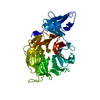
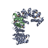
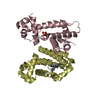
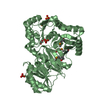
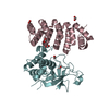
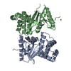
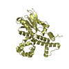
| Title | Crystal structure of Nas2 N-terminal domain complexed with PAN-Rpt5C chimera | ||||||
 Components Components |
| ||||||
 Keywords Keywords | HYDROLASE/CHAPERONE /  Four-helix bundle / Proteasome ATPase subunit / Proteasome assembly chaperone / ATP Binding / HYDROLASE-CHAPERONE complex Four-helix bundle / Proteasome ATPase subunit / Proteasome assembly chaperone / ATP Binding / HYDROLASE-CHAPERONE complex | ||||||
| Function / homology |  Function and homology information Function and homology informationproteasome-activating nucleotidase complex / Regulation of PTEN stability and activity / KEAP1-NFE2L2 pathway / CDK-mediated phosphorylation and removal of Cdc6 / Neddylation / FBXL7 down-regulates AURKA during mitotic entry and in early mitosis / Ubiquitin Mediated Degradation of Phosphorylated Cdc25A / Orc1 removal from chromatin / MAPK6/MAPK4 signaling / proteasome regulatory particle assembly ...proteasome-activating nucleotidase complex / Regulation of PTEN stability and activity / KEAP1-NFE2L2 pathway / CDK-mediated phosphorylation and removal of Cdc6 / Neddylation / FBXL7 down-regulates AURKA during mitotic entry and in early mitosis / Ubiquitin Mediated Degradation of Phosphorylated Cdc25A / Orc1 removal from chromatin / MAPK6/MAPK4 signaling / proteasome regulatory particle assembly / Antigen processing: Ubiquitination & Proteasome degradation / proteasome-activating activity / proteasome regulatory particle, base subcomplex / Cross-presentation of soluble exogenous antigens (endosomes) / TNFR2 non-canonical NF-kB pathway / protein unfolding / Ub-specific processing proteases / positive regulation of RNA polymerase II transcription preinitiation complex assembly / Neutrophil degranulation / proteasome complex / proteasomal protein catabolic process / ubiquitin-dependent protein catabolic process / proteasome-mediated ubiquitin-dependent protein catabolic process / ATP hydrolysis activity / ATP binding / nucleus / cytosol / cytoplasmSimilarity search - Function | ||||||
| Biological species |   Pyrococcus furiosus (archaea) Pyrococcus furiosus (archaea) Saccharomyces cerevisiae (brewer's yeast) Saccharomyces cerevisiae (brewer's yeast) | ||||||
| Method | X-RAY DIFFRACTION / SYNCHROTRON / MOLECULAR REPLACEMENT / Resolution: 4 Å | ||||||
 Authors Authors | Satoh, T. / Saeki, Y. / Hiromoto, T. / Wang, Y.-H. / Uekusa, Y. / Yagi, H. / Yoshihara, H. / Yagi-Utsumi, M. / Mizushima, T. / Tanaka, K. / Kato, K. | ||||||
 Citation Citation | Journal: Structure / Year: 2014 Title: Structural basis for proteasome formation controlled by an assembly chaperone nas2. Authors: Satoh, T. / Saeki, Y. / Hiromoto, T. / Wang, Y.H. / Uekusa, Y. / Yagi, H. / Yoshihara, H. / Yagi-Utsumi, M. / Mizushima, T. / Tanaka, K. / Kato, K. | ||||||
| History |
|
- Structure visualization
| Structure viewer | Molecule: MolmilJmol/JSmol |
|---|
- Downloads & links
-Download
| PDBx/mmCIF format | 3whl.cif.gz | 267.1 KB | Display | PDBx/mmCIF format |
|---|---|---|---|---|
| PDB format | pdb3whl.ent.gz | 215.2 KB | Display | PDB format |
| PDBx/mmJSON format | 3whl.json.gz | Tree view | PDBx/mmJSON format | |
| Others |  Other downloads Other downloads |
-Validation report
| Arichive directory | https://data.pdbj.org/pub/pdb/validation_reports/wh/3whlftp://data.pdbj.org/pub/pdb/validation_reports/wh/3whl | HTTPS FTP |
|---|
-Related structure data
| Related structure data |  3whjC 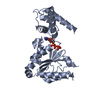 3whkSC C: citing same article ( S: Starting model for refinement |
|---|---|
| Similar structure data |


-Links
 PDBj
PDBj


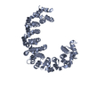
- Assembly
| Deposited unit | 
| ||||||||
|---|---|---|---|---|---|---|---|---|---|
| 1 | 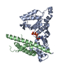
| ||||||||
| 2 | 
| ||||||||
| 3 | 
| ||||||||
| 4 | 
| ||||||||
| Unit cell |
|
-Components
| #1: Protein | Mass: 30023.271 Da / Num. of mol.: 4 Source method: isolated from a genetically manipulated source Details: Fusion protein of residues 125-309 FROM Proteasome-activating nucleotidase (PAN, UNP Q8U4H3), linker (EF), and residues 356-434 from 26S protease regulatory subunit 6A (Rpt5, UNP P33297). Source: (gene. exp.) Pyrococcus furiosus (archaea), (gene. exp.) Saccharomyces cerevisiae (brewer's yeast)Strain: DSM 3638, S288c / Gene: pan, PF0115, O3258, RPT5, YOR117W, YOR3258W, YTA1 / Plasmid: pET-28b / Production host:  Escherichia coli (E. coli) / Strain (production host): BL21-CodonPlus(DE3) / References: UniProt: Q8U4H3, UniProt: P33297 Escherichia coli (E. coli) / Strain (production host): BL21-CodonPlus(DE3) / References: UniProt: Q8U4H3, UniProt: P33297#2: Protein | Mass: 13861.723 Da / Num. of mol.: 4 / Fragment: N-terminal domain, UNP residues 1-120 Source method: isolated from a genetically manipulated source Source: (gene. exp.) Saccharomyces cerevisiae (brewer's yeast)Strain: ATCC 204508 / S288c / Gene: NAS2, YIL007C / Plasmid: pCold-MBP / Production host: Escherichia coli (E. coli) / Strain (production host): BL21-CodonPlus(DE3) / References: UniProt: P40555#3: Chemical | ChemComp-ATP / Adenosine triphosphate  Mass: 507.181 Da / Num. of mol.: 4 / Source method: obtained synthetically / Formula: C10H16N5O13P3 / Comment: ATP, energy-carrying molecule*YM Mass: 507.181 Da / Num. of mol.: 4 / Source method: obtained synthetically / Formula: C10H16N5O13P3 / Comment: ATP, energy-carrying molecule*YM |
|---|
-Experimental details
-Experiment
| Experiment | Method: X-RAY DIFFRACTION / Number of used crystals: 1 |
|---|
- Sample preparation
| Crystal | Density Matthews: 2.54 Å3/Da / Density % sol: 51.49 % |
|---|---|
| Crystal grow | Temperature: 293 K / Method: vapor diffusion, sitting drop / pH: 6 Details: 8% PEG 6000, 0.1M MES (pH6.0), 80mM MgCl2, VAPOR DIFFUSION, SITTING DROP, temperature 293K |
-Data collection
| Diffraction | Mean temperature: 100 K |
|---|---|
| Diffraction source | Source: SYNCHROTRON / Site: SPring-8  / Beamline: BL44XU / Wavelength: 0.9 Å / Beamline: BL44XU / Wavelength: 0.9 Å |
| Detector | Type: RAYONIX MX-225 / Detector: CCD / Date: Jan 30, 2012 |
| Radiation | Monochromator: Si 111 / Protocol: SINGLE WAVELENGTH / Monochromatic (M) / Laue (L): M / Scattering type: x-ray |
| Radiation wavelength | Wavelength: 0.9 Å / Relative weight: 1 |
| Reflection | Resolution: 4→50 Å / Num. all: 15872 / Num. obs: 15849 / % possible obs: 100 % / Observed criterion σ(I): -3 / Redundancy: 5.5 % / Biso Wilson estimate: 110.8 Å2 / Rmerge(I) obs: 0.1 / Net I/σ(I): 28.1 |
| Reflection shell | Resolution: 4→4.07 Å / Redundancy: 5.5 % / Rmerge(I) obs: 0.554 / Mean I/σ(I) obs: 5 / Num. unique all: 787 / % possible all: 100 |
- Processing
| Software |
| ||||||||||||||||||||||||||||||||||||
|---|---|---|---|---|---|---|---|---|---|---|---|---|---|---|---|---|---|---|---|---|---|---|---|---|---|---|---|---|---|---|---|---|---|---|---|---|---|
| Refinement | Method to determine structure: MOLECULAR REPLACEMENT Starting model: 3WHK Resolution: 4→19.94 Å / Occupancy max: 1 / Occupancy min: 1 / FOM work R set: 0.764 / SU ML: 0.47 / σ(F): 0.65 / Phase error: 29.39 / Stereochemistry target values: ML
| ||||||||||||||||||||||||||||||||||||
| Solvent computation | Shrinkage radii: 0.9 Å / VDW probe radii: 1.11 Å / Solvent model: FLAT BULK SOLVENT MODEL | ||||||||||||||||||||||||||||||||||||
| Displacement parameters | Biso max: 248.44 Å2 / Biso mean: 148.2997 Å2 / Biso min: 90.23 Å2 | ||||||||||||||||||||||||||||||||||||
| Refinement step | Cycle: LAST / Resolution: 4→19.94 Å
| ||||||||||||||||||||||||||||||||||||
| Refine LS restraints |
| ||||||||||||||||||||||||||||||||||||
| LS refinement shell | Refine-ID: X-RAY DIFFRACTION / Total num. of bins used: 5
|
