 Movie
Movie Controller

[English] 日本語
 Yorodumi
- PDB-2dzn: Crystal structure analysis of yeast Nas6p complexed with the prot... -
+ Open data

- Basic information
| Entry | Database: PDB / ID: 2dzn | ||||||
|---|---|---|---|---|---|---|---|
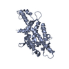
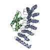
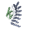
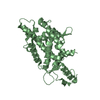
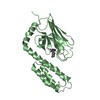
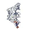
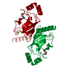
| Title | Crystal structure analysis of yeast Nas6p complexed with the proteasome subunit, rpt3 | ||||||
 Components Components |
| ||||||
 Keywords Keywords |  PROTEIN BINDING / ankyrin repeats / a-helical domain / Structural Genomics / NPPSFA / National Project on Protein Structural and Functional Analyses / RIKEN Structural Genomics/Proteomics Initiative / RSGI PROTEIN BINDING / ankyrin repeats / a-helical domain / Structural Genomics / NPPSFA / National Project on Protein Structural and Functional Analyses / RIKEN Structural Genomics/Proteomics Initiative / RSGI | ||||||
| Function / homology |  Function and homology information Function and homology informationproteasome regulatory particle binding / proteasome regulatory particle assembly / proteasome-activating activity / proteasome regulatory particle, base subcomplex / Cross-presentation of soluble exogenous antigens (endosomes) / TNFR2 non-canonical NF-kB pathway / Ub-specific processing proteases / positive regulation of RNA polymerase II transcription preinitiation complex assembly / proteasome complex / ubiquitin-dependent protein catabolic process ...proteasome regulatory particle binding / proteasome regulatory particle assembly / proteasome-activating activity / proteasome regulatory particle, base subcomplex / Cross-presentation of soluble exogenous antigens (endosomes) / TNFR2 non-canonical NF-kB pathway / Ub-specific processing proteases / positive regulation of RNA polymerase II transcription preinitiation complex assembly / proteasome complex / ubiquitin-dependent protein catabolic process / proteasome-mediated ubiquitin-dependent protein catabolic process / ATP hydrolysis activity / ATP binding / identical protein binding / nucleus / cytosol / cytoplasmSimilarity search - Function | ||||||
| Biological species |  Saccharomyces cerevisiae (brewer's yeast) Saccharomyces cerevisiae (brewer's yeast) | ||||||
| Method | X-RAY DIFFRACTION / SYNCHROTRON / MOLECULAR REPLACEMENT / Resolution: 2.2 Å | ||||||
 Authors Authors | Yokoyama, S. / Padmanabhan, B. / RIKEN Structural Genomics/Proteomics Initiative (RSGI) | ||||||
 Citation Citation | Journal: Biochem.Biophys.Res.Commun. / Year: 2007 Title: Structural basis for the recognition between the regulatory particles Nas6 and Rpt3 of the yeast 26S proteasome Authors: Nakamura, Y. / Umehara, T. / Tanaka, A. / Horikoshi, M. / Padmanabhan, B. / Yokoyama, S. | ||||||
| History |
|
- Structure visualization
| Structure viewer | Molecule: MolmilJmol/JSmol |
|---|
- Downloads & links
-Download
| PDBx/mmCIF format | 2dzn.cif.gz | 187.4 KB | Display | PDBx/mmCIF format |
|---|---|---|---|---|
| PDB format | pdb2dzn.ent.gz | 150.5 KB | Display | PDB format |
| PDBx/mmJSON format | 2dzn.json.gz | Tree view | PDBx/mmJSON format | |
| Others |  Other downloads Other downloads |
-Validation report
| Arichive directory | https://data.pdbj.org/pub/pdb/validation_reports/dz/2dznftp://data.pdbj.org/pub/pdb/validation_reports/dz/2dzn | HTTPS FTP |
|---|
-Related structure data
| Related structure data | |
|---|---|
| Similar structure data | |
| Other databases |


-Links
 PDBj
PDBj


- Assembly
| Deposited unit | 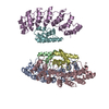
| ||||||||
|---|---|---|---|---|---|---|---|---|---|
| 1 | 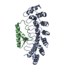
| ||||||||
| 2 | 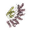
| ||||||||
| 3 | 
| ||||||||
| Unit cell |
|
-Components
| #1: Protein | Mass: 25648.268 Da / Num. of mol.: 3 Source method: isolated from a genetically manipulated source Source: (gene. exp.) Saccharomyces cerevisiae (brewer's yeast)Plasmid: PETDUET1 / Production host:  Escherichia coli (E. coli) / References: UniProt: P50086 Escherichia coli (E. coli) / References: UniProt: P50086#2: Protein | Mass: 9208.535 Da / Num. of mol.: 3 / Fragment: C-terminal domain Source method: isolated from a genetically manipulated source Source: (gene. exp.) Saccharomyces cerevisiae (brewer's yeast)Plasmid: PETDUET1 / Production host: Escherichia coli (E. coli) / References: UniProt: P33298#3: Water | ChemComp-HOH / | Water Mass: 18.015 Da / Num. of mol.: 472 / Source method: isolated from a natural source / Formula: H2O Mass: 18.015 Da / Num. of mol.: 472 / Source method: isolated from a natural source / Formula: H2O |
|---|
-Experimental details
-Experiment
| Experiment | Method: X-RAY DIFFRACTION / Number of used crystals: 1 |
|---|
- Sample preparation
| Crystal | Density Matthews: 2.08 Å3/Da / Density % sol: 40.9 % |
|---|---|
| Crystal grow | Temperature: 298 K / Method: vapor diffusion / pH: 6.5 Details: PEG6K, MES, pH 6.50, VAPOR DIFFUSION, temperature 298K |
-Data collection
| Diffraction | Mean temperature: 120 K |
|---|---|
| Diffraction source | Source: SYNCHROTRON / Site: Photon Factory  / Beamline: AR-NW12A / Wavelength: 1 / Wavelength: 1 Å / Beamline: AR-NW12A / Wavelength: 1 / Wavelength: 1 Å |
| Detector | Type: ADSC QUANTUM 4 / Detector: CCD / Date: May 20, 2005 / Details: MIRRORS |
| Radiation | Protocol: SINGLE WAVELENGTH / Monochromatic (M) / Laue (L): M / Scattering type: x-ray |
| Radiation wavelength | Wavelength: 1 Å / Relative weight: 1 |
| Reflection | Resolution: 2.2→50 Å / Num. obs: 43936 / % possible obs: 99.6 % / Observed criterion σ(I): -3 / Redundancy: 3.7 % / Rmerge(I) obs: 0.056 |
| Reflection shell | Resolution: 2.2→2.28 Å / Redundancy: 3.8 % / Rmerge(I) obs: 0.203 / % possible all: 100 |
- Processing
| Software |
| ||||||||||||||||||||
|---|---|---|---|---|---|---|---|---|---|---|---|---|---|---|---|---|---|---|---|---|---|
| Refinement | Method to determine structure: MOLECULAR REPLACEMENT Starting model: GANKYRIN Resolution: 2.2→20 Å / Isotropic thermal model: ISOTROPIC / Cross valid method: THROUGHOUT / σ(F): 0 / Stereochemistry target values: Engh & Huber
| ||||||||||||||||||||
| Displacement parameters | Biso mean: 42.16 Å2 | ||||||||||||||||||||
| Refinement step | Cycle: LAST / Resolution: 2.2→20 Å
| ||||||||||||||||||||
| Refine LS restraints |
| ||||||||||||||||||||
| LS refinement shell | Resolution: 2.2→2.26 Å /
|
