 Movie
Movie Controller


+ Open data

- Basic information
| Entry | Database: PDB / ID: 3vh7 | ||||||
|---|---|---|---|---|---|---|---|
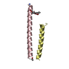
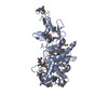
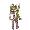
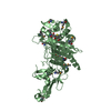
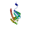
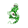
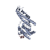
| Title | Structure of HIV-1 gp41 NHR/fusion inhibitor complex P21 | ||||||
 Components Components |
| ||||||
 Keywords Keywords |  MEMBRANE PROTEIN/INHIBITOR / 6-helix bundle / membrane fusion inhibition / MEMBRANE PROTEIN-INHIBITOR complex MEMBRANE PROTEIN/INHIBITOR / 6-helix bundle / membrane fusion inhibition / MEMBRANE PROTEIN-INHIBITOR complex | ||||||
| Function / homology |  Function and homology information Function and homology informationhost cell periphery / CD4 receptor binding / Dectin-2 family / positive regulation of plasma membrane raft polarization / positive regulation of receptor clustering / positive regulation of establishment of T cell polarity / virus-mediated perturbation of host defense response / host cell endosome membrane / clathrin-dependent endocytosis of virus by host cell / host cell perinuclear region of cytoplasm ...host cell periphery / CD4 receptor binding / Dectin-2 family / positive regulation of plasma membrane raft polarization / positive regulation of receptor clustering / positive regulation of establishment of T cell polarity / virus-mediated perturbation of host defense response / host cell endosome membrane / clathrin-dependent endocytosis of virus by host cell / host cell perinuclear region of cytoplasm / viral protein processing / fusion of virus membrane with host plasma membrane / fusion of virus membrane with host endosome membrane / viral envelope / protein-containing complex binding / virion attachment to host cell / host cell plasma membrane / virion membrane / structural molecule activity / membraneSimilarity search - Function | ||||||
| Biological species |   Human immunodeficiency virus type 1 Human immunodeficiency virus type 1 | ||||||
| Method | X-RAY DIFFRACTION / SYNCHROTRON / MOLECULAR REPLACEMENT / Resolution: 2.019 Å | ||||||
 Authors Authors | Yao, X. / Waltersperger, S. / Wang, M.T. / Cui, S. | ||||||
 Citation Citation | Journal: J.Biol.Chem. / Year: 2012 Title: Structural basis of potent and broad HIV-1 fusion inhibitor CP32M Authors: Yao, X. / Chong, H. / Zhang, C. / Qiu, Z. / Qin, B. / Han, R. / Waltersperger, S. / Wang, M.T. / He, Y. / Cui, S. | ||||||
| History |
|
- Structure visualization
| Structure viewer | Molecule: MolmilJmol/JSmol |
|---|
- Downloads & links
-Download
| PDBx/mmCIF format | 3vh7.cif.gz | 110 KB | Display | PDBx/mmCIF format |
|---|---|---|---|---|
| PDB format | pdb3vh7.ent.gz | 87.3 KB | Display | PDB format |
| PDBx/mmJSON format | 3vh7.json.gz | Tree view | PDBx/mmJSON format | |
| Others |  Other downloads Other downloads |
-Validation report
| Arichive directory | https://data.pdbj.org/pub/pdb/validation_reports/vh/3vh7ftp://data.pdbj.org/pub/pdb/validation_reports/vh/3vh7 | HTTPS FTP |
|---|
-Related structure data
| Related structure data |  3vgyC 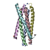 3f4yS C: citing same article ( S: Starting model for refinement |
|---|---|
| Similar structure data |


-Links
 PDBj
PDBj


- Assembly
| Deposited unit | 
| ||||||||
|---|---|---|---|---|---|---|---|---|---|
| 1 |
| ||||||||
| Unit cell |
|
-Components
| #1: Protein | Mass: 6413.350 Da / Num. of mol.: 3 / Fragment: NHR (UNP RESIDUES (546-588) / Source method: obtained synthetically / Details: This sequence occurs naturally. / Source: (synth.) Human immunodeficiency virus type 1 / References: UniProt: P03375#2: Protein/peptide | Mass: 4295.733 Da / Num. of mol.: 3 / Source method: obtained synthetically Details: This sequence does not occur naturally, but designed based on sequence of HIV-1 gp41 CHR 621-652. #3: Chemical | ChemComp-MG / |   Mass: 24.305 Da / Num. of mol.: 1 / Source method: obtained synthetically / Formula: Mg Mass: 24.305 Da / Num. of mol.: 1 / Source method: obtained synthetically / Formula: Mg#4: Water | ChemComp-HOH / | Water Mass: 18.015 Da / Num. of mol.: 68 / Source method: isolated from a natural source / Formula: H2O Mass: 18.015 Da / Num. of mol.: 68 / Source method: isolated from a natural source / Formula: H2O |
|---|
-Experimental details
-Experiment
| Experiment | Method: X-RAY DIFFRACTION / Number of used crystals: 1 |
|---|
- Sample preparation
| Crystal | Density Matthews: 1.89 Å3/Da / Density % sol: 34.99 % Description: the entry contains Friedel pairs in F_Plus/Minus columns |
|---|---|
| Crystal grow | Temperature: 295 K / Method: vapor diffusion, hanging drop / pH: 4.6 Details: 0.1M MgCl2, 0.1M Sodium acetate, 25% PEG 400, pH 4.6, VAPOR DIFFUSION, HANGING DROP, temperature 295K |
-Data collection
| Diffraction | Mean temperature: 100 K |
|---|---|
| Diffraction source | Source: SYNCHROTRON / Site: SLS  / Beamline: X06DA / Wavelength: 1 Å / Beamline: X06DA / Wavelength: 1 Å |
| Detector | Type: MARMOSAIC 225 mm CCD / Detector: CCD / Date: Mar 14, 2011 |
| Radiation | Monochromator: Bartels Monochromator Crystal Type Si (111) / Protocol: SINGLE WAVELENGTH / Monochromatic (M) / Laue (L): M / Scattering type: x-ray |
| Radiation wavelength | Wavelength: 1 Å / Relative weight: 1 |
| Reflection | Resolution: 2.019→42.607 Å / Num. obs: 14977 / % possible obs: 90.8 % / Observed criterion σ(F): 3 / Observed criterion σ(I): 3 / Redundancy: 1.97 % / Rmerge(I) obs: 0.084 / Rsym value: 0.085 / Net I/σ(I): 7.8 |
| Reflection shell | Resolution: 2.02→2.14 Å / Redundancy: 1.24 % / Rmerge(I) obs: 0.747 / Mean I/σ(I) obs: 1.18 / Num. unique all: 3185 / Rsym value: 0.764 / % possible all: 63.3 |
- Processing
| Software |
| ||||||||||||||||||||||||||||||||||||||||||
|---|---|---|---|---|---|---|---|---|---|---|---|---|---|---|---|---|---|---|---|---|---|---|---|---|---|---|---|---|---|---|---|---|---|---|---|---|---|---|---|---|---|---|---|
| Refinement | Method to determine structure: MOLECULAR REPLACEMENT Starting model: PDB ID 3F4Y Resolution: 2.019→42.607 Å / SU ML: 0.3 / Cross valid method: THROUGHOUT / σ(F): 1.45 / Phase error: 28.02 / Stereochemistry target values: ML Details: the entry contains Friedel pairs in F_Plus/Minus columns
| ||||||||||||||||||||||||||||||||||||||||||
| Solvent computation | Shrinkage radii: 0.86 Å / VDW probe radii: 1.1 Å / Solvent model: FLAT BULK SOLVENT MODEL / Bsol: 55.303 Å2 / ksol: 0.385 e/Å3 | ||||||||||||||||||||||||||||||||||||||||||
| Displacement parameters |
| ||||||||||||||||||||||||||||||||||||||||||
| Refinement step | Cycle: LAST / Resolution: 2.019→42.607 Å
| ||||||||||||||||||||||||||||||||||||||||||
| Refine LS restraints |
| ||||||||||||||||||||||||||||||||||||||||||
| LS refinement shell |
|
