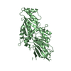
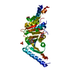
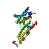
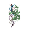
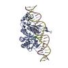
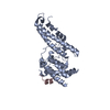
- PDB-4nuu: Heterotrimer structure of Region II from Plasmodium vivax Duffy B... -
+
Open data
ID or keywords:
Loading...
-
Basic information
Entry
Database: PDB / ID: 4nuu
Title
Heterotrimer structure of Region II from Plasmodium vivax Duffy Binding Protein (PvDBP) bound to the ectodomain of the Duffy Antigen Receptor for Chemokines (DARC)
Components
Duffy antigen/chemokine receptor
Duffy receptor
Keywords
membrane protein / cell invasion / Duffy Binding Like (DBL) Domain Fold / GPCR / Adhesion / Invasion / Red blood cell binding / Chemokine Binding / Duffy Antigen Receptor for Chemokines / Membrane / protein binding
Function / homology
Function and homology information
regulation of chemokine production / C-C chemokine binding / chemokine-mediated signaling pathway / Peptide ligand-binding receptors / G protein-coupled receptor activity / recycling endosome / defense response / transmembrane signaling receptor activity / signaling receptor activity / early endosome ...regulation of chemokine production / C-C chemokine binding / chemokine-mediated signaling pathway / Peptide ligand-binding receptors / G protein-coupled receptor activity / recycling endosome / defense response / transmembrane signaling receptor activity / signaling receptor activity / early endosome / host cell surface receptor binding / inflammatory response / membrane / identical protein binding / plasma membrane Similarity search - Function
In the structure databanks used in Yorodumi, some data are registered as the other names, "COVID-19 virus" and "2019-nCoV". Here are the details of the virus and the list of structure data.
Jan 31, 2019. EMDB accession codes are about to change! (news from PDBe EMDB page)
EMDB accession codes are about to change! (news from PDBe EMDB page)
The allocation of 4 digits for EMDB accession codes will soon come to an end. Whilst these codes will remain in use, new EMDB accession codes will include an additional digit and will expand incrementally as the available range of codes is exhausted. The current 4-digit format prefixed with “EMD-” (i.e. EMD-XXXX) will advance to a 5-digit format (i.e. EMD-XXXXX), and so on. It is currently estimated that the 4-digit codes will be depleted around Spring 2019, at which point the 5-digit format will come into force.
The EM Navigator/Yorodumi systems omit the EMD- prefix.
Related info.:Q: What is EMD? / ID/Accession-code notation in Yorodumi/EM Navigator
Yorodumi is a browser for structure data from EMDB, PDB, SASBDB, etc.
This page is also the successor to EM Navigator detail page, and also detail information page/front-end page for Omokage search.
The word "yorodu" (or yorozu) is an old Japanese word meaning "ten thousand". "mi" (miru) is to see.
Related info.:EMDB / PDB / SASBDB / Comparison of 3 databanks / Yorodumi Search / Aug 31, 2016. New EM Navigator & Yorodumi / Yorodumi Papers / Jmol/JSmol / Function and homology information / Changes in new EM Navigator and Yorodumi
 Movie
Movie Controller

 Yorodumi
 Open data
 Basic information
 Components
 Keywords
 membrane protein / cell invasion / Duffy Binding Like (DBL) Domain Fold /
  Function and homology information

 Authors
 Citation
 Structure visualization
 Downloads & links
 Other downloads
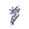


 PDBj
PDBj


 Assembly


 Mass: 18.015 Da / Num. of mol.: 462 / Source method: isolated from a natural source / Formula: H2O
 Sample preparation
 / Beamline: 4.2.2 / Wavelength: 0.9792909 Å
 Processing