[English] 日本語
Yorodumi
- PDB-4r2j: Crystal structure of YdaA (Universal Stress Protein E) from Salmo... -

+
Open data


ID or keywords:

Loading...

-
Basic information

Entry
Database: PDB / ID: 4r2j
TitleCrystal structure of YdaA (Universal Stress Protein E) from Salmonella typhimurium
ComponentsUniversal stress protein E
KeywordsMETAL BINDING PROTEIN / UNKNOWN FUNCTION / 
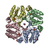
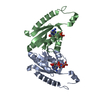
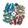
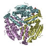
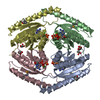
Universal stress protein / HUP domain / Internal Symmetry / Stress tolerance / ATP binding
Function / homology
Function and homology information


Rossmann fold - #12370 / UspA / Universal stress protein family / Rossmann fold / 3-Layer(aba) Sandwich / Alpha Beta
Similarity search - Domain/homology
alpha-D-glucopyranose / PHOSPHATE ION / Universal stress protein E
Similarity search - Component
Biological speciesSalmonella enterica subsp. enterica serovar Typhimurium str. LT2 (bacteria)
MethodX-RAY DIFFRACTION / SYNCHROTRON / MOLECULAR REPLACEMENT / Resolution: 2.36 Å
AuthorsBangera, M. / Murthy, M.R.N.
CitationJournal: J.Struct.Biol. / Year: 2015
Title: Structural and functional analysis of two universal stress proteins YdaA and YnaF from Salmonella typhimurium: possible roles in microbial stress tolerance.
Authors: Bangera, M. / Panigrahi, R. / Sagurthi, S.R. / Savithri, H.S. / Murthy, M.R.
History
DepositionAug 12, 2014Deposition site: RCSB / Processing site: PDBJ
Revision 1.0Feb 4, 2015Provider: repository / Type: Initial release
Revision 1.1Jul 29, 2020Group: Data collection / Database references ...Data collection / Database references / Derived calculations / Structure summary
Category: chem_comp / entity ...chem_comp / entity / pdbx_chem_comp_identifier / pdbx_entity_nonpoly / pdbx_struct_conn_angle / struct_conn / struct_ref_seq_dif / struct_site / struct_site_gen
Item: _chem_comp.name / _chem_comp.type ..._chem_comp.name / _chem_comp.type / _entity.pdbx_description / _pdbx_entity_nonpoly.name / _pdbx_struct_conn_angle.ptnr1_auth_seq_id / _pdbx_struct_conn_angle.ptnr1_label_atom_id / _pdbx_struct_conn_angle.ptnr1_label_seq_id / _pdbx_struct_conn_angle.ptnr3_auth_seq_id / _pdbx_struct_conn_angle.ptnr3_label_atom_id / _pdbx_struct_conn_angle.ptnr3_label_seq_id / _pdbx_struct_conn_angle.value / _struct_conn.pdbx_dist_value / _struct_conn.ptnr1_auth_seq_id / _struct_conn.ptnr1_label_atom_id / _struct_conn.ptnr1_label_seq_id / _struct_ref_seq_dif.details
Description: Carbohydrate remediation / Provider: repository / Type: Remediation
Revision 1.2Aug 24, 2022Group: Database references / Structure summary
Category: chem_comp / citation ...chem_comp / citation / citation_author / database_2
Item: _chem_comp.pdbx_synonyms / _citation.journal_volume ..._chem_comp.pdbx_synonyms / _citation.journal_volume / _citation.page_first / _citation.page_last / _citation.title / _citation_author.name / _database_2.pdbx_DOI / _database_2.pdbx_database_accession
Revision 1.3Nov 8, 2023Group: Data collection / Refinement description
Category: chem_comp_atom / chem_comp_bond / pdbx_initial_refinement_model

-
Structure visualization

Structure viewerMolecule:
MolmilJmol/JSmol

Downloads & links

-
Assembly

Deposited unit
A: Universal stress protein E
hetero molecules


Theoretical massNumber of molelcules
Total (without water)37,8716
Polymers37,2891
Non-polymers5835
Water61334
1
A: Universal stress protein E
hetero molecules

A: Universal stress protein E
hetero molecules


Theoretical massNumber of molelcules
Total (without water)75,74312
Polymers74,5772
Non-polymers1,16610
Water362
TypeNameSymmetry operationNumber
identity operation1_555x,y,z1
crystal symmetry operation4_555y,x,-z1
Buried area3310 Å2
ΔGint-76 kcal/mol
Surface area26630 Å2
MethodPISA
Unit cell
Length a, b, c (Å)85.770, 85.770, 74.991
Angle α, β, γ (deg.)90.00, 90.00, 120.00
Int Tables number152
Space group name H-MP3121

-
Components

-
Protein / Sugars , 2 types, 3 molecules A

#1: Protein Universal stress protein E


Mass: 37288.547 Da / Num. of mol.: 1
Source method: isolated from a genetically manipulated source
Source: (gene. exp.) Salmonella enterica subsp. enterica serovar Typhimurium str. LT2 (bacteria)
Strain: LT2 / Gene: STM1661, uspE, ydaA uspE STM1661 / Plasmid: pRSET C / Production host: Escherichia coli (E. coli) / Strain (production host): BL21(DE3)pLysS / References: UniProt: Q8ZP84
#5: Sugar ChemComp-GLC / alpha-D-glucopyranose / alpha-D-glucose / D-glucose / glucose / Glucose


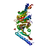
Type: D-saccharide, alpha linking / Mass: 180.156 Da / Num. of mol.: 2
Source method: isolated from a genetically manipulated source
Formula: C6H12O6
IdentifierTypeProgram
DGlcpaCONDENSED IUPAC CARBOHYDRATE SYMBOLGMML 1.0
a-D-glucopyranoseCOMMON NAMEGMML 1.0
a-D-GlcpIUPAC CARBOHYDRATE SYMBOLPDB-CARE 1.0
GlcSNFG CARBOHYDRATE SYMBOLGMML 1.0

-
Non-polymers , 4 types, 37 molecules

#2: Chemical ChemComp-ZN / ZINC ION


Mass: 65.409 Da / Num. of mol.: 1 / Source method: obtained synthetically / Formula: Zn
#3: Chemical ChemComp-PO4 / PHOSPHATE ION / Phosphate


Mass: 94.971 Da / Num. of mol.: 1 / Source method: obtained synthetically / Formula: PO4
#4: Chemical ChemComp-EDO / 1,2-ETHANEDIOL / ETHYLENE GLYCOL / Ethylene glycol


Mass: 62.068 Da / Num. of mol.: 1 / Source method: obtained synthetically / Formula: C2H6O2
#6: Water ChemComp-HOH / water / Water


Mass: 18.015 Da / Num. of mol.: 34 / Source method: isolated from a natural source / Formula: H2O

-
Experimental details

-
Experiment

ExperimentMethod: X-RAY DIFFRACTION / Number of used crystals: 1

-
Sample preparation

CrystalDensity Matthews: 2.14 Å3/Da / Density % sol: 42.4 %
Crystal growTemperature: 298 K / Method: under oil, microbatch / pH: 5.8
Details: 0.2M Magnesium Nitrate hexahydrate, 20% PEG 3350, 10mM Adenosine 5'-(beta,gamma-imido)triphosphate, 0.2% n-dodecyl beta-D-maltoside, pH 5.8, Under oil, Microbatch, temperature 298.0K

-
Data collection

DiffractionMean temperature: 100 K
Diffraction sourceSource: SYNCHROTRON / Site: ESRF / Beamline: BM14 / Wavelength: 0.97625 Å
DetectorType: MARMOSAIC 225 mm CCD / Detector: CCD / Date: Feb 14, 2011 / Details: bent collimating mirror and toroid
RadiationMonochromator: Si(111) monochromator / Protocol: SINGLE WAVELENGTH / Monochromatic (M) / Laue (L): M / Scattering type: x-ray
Radiation wavelengthWavelength: 0.97625 Å / Relative weight: 1
ReflectionResolution: 2.36→42.88 Å / Num. obs: 13536 / % possible obs: 99.8 % / Redundancy: 5.7 % / Biso Wilson estimate: 45.2 Å2 / Rsym value: 0.119
Reflection shellResolution: 2.36→2.48 Å / Redundancy: 5.5 % / Mean I/σ(I) obs: 2.2 / Num. unique all: 1929 / Rsym value: 0.642 / % possible all: 98.9

-
Processing

Software
NameVersionClassification
MxCuBE2data collection
PHASERphasing
REFMAC5.8.0049refinement
MOSFLMdata reduction
SCALAdata scaling
RefinementMethod to determine structure: MOLECULAR REPLACEMENT
Starting model: PDB ID 3OLQ
Resolution: 2.36→37.14 Å / Cor.coef. Fo:Fc: 0.951 / Cor.coef. Fo:Fc free: 0.933 / SU B: 20.386 / SU ML: 0.235 / Cross valid method: THROUGHOUT / ESU R: 0.37 / ESU R Free: 0.259 / Stereochemistry target values: MAXIMUM LIKELIHOOD / Details: HYDROGENS HAVE BEEN USED IF PRESENT IN THE INPUT
RfactorNum. reflection% reflectionSelection details
Rfree0.26186 667 4.9 %RANDOM
Rwork0.21193 ---
obs0.21435 12849 99.79 %-
Solvent computationIon probe radii: 0.8 Å / Shrinkage radii: 0.8 Å / VDW probe radii: 1.2 Å / Solvent model: MASK
Displacement parametersBiso mean: 59.55 Å2
Baniso -1Baniso -2Baniso -3
1-1.15 Å20.57 Å2-0 Å2
2--1.15 Å2-0 Å2
3----3.72 Å2
Refinement stepCycle: LAST / Resolution: 2.36→37.14 Å
ProteinNucleic acidLigandSolventTotal
Num. atoms2115 0 34 34 2183
Refine LS restraints
Refine-IDTypeDev idealDev ideal targetNumber
X-RAY DIFFRACTIONr_bond_refined_d0.0130.0192192
X-RAY DIFFRACTIONr_angle_refined_deg1.7341.9663001
X-RAY DIFFRACTIONr_dihedral_angle_1_deg6.6735280
X-RAY DIFFRACTIONr_dihedral_angle_2_deg38.78424.58885
X-RAY DIFFRACTIONr_dihedral_angle_3_deg17.71215311
X-RAY DIFFRACTIONr_dihedral_angle_4_deg18.201159
X-RAY DIFFRACTIONr_chiral_restr0.1020.2364
X-RAY DIFFRACTIONr_gen_planes_refined0.0080.0211628
X-RAY DIFFRACTIONr_mcbond_it2.6783.9621135
X-RAY DIFFRACTIONr_mcangle_it3.9515.9231410
X-RAY DIFFRACTIONr_scbond_it3.8764.2381057
X-RAY DIFFRACTIONr_long_range_B_refined7.26334.2513270
LS refinement shellResolution: 2.356→2.417 Å / Total num. of bins used: 20
RfactorNum. reflection% reflection
Rfree0.268 46 -
Rwork0.286 913 -
obs--97.66 %
Refinement TLS params.Method: refined / Origin x: 25.405 Å / Origin y: 16.474 Å / Origin z: 6.161 Å
111213212223313233
T0.0991 Å20.0238 Å2-0.0514 Å2-0.0267 Å20.0092 Å2--0.0689 Å2
L1.4166 °2-0.4553 °2-0.6366 °2-2.1973 °20.486 °2--6.5914 °2
S0.0708 Å °-0.0436 Å °-0.0478 Å °-0.0941 Å °-0.0055 Å °0.0883 Å °0.3938 Å °0.0236 Å °-0.0653 Å °

+
About Yorodumi

-
News

-
Feb 9, 2022. New format data for meta-information of EMDB entries

New format data for meta-information of EMDB entries

  • Version 3 of the EMDB header file is now the official format.
  • The previous official version 1.9 will be removed from the archive.

Related info.:EMDB header

External links:wwPDB to switch to version 3 of the EMDB data model

-
Aug 12, 2020. Covid-19 info

Covid-19 info

URL: https://pdbj.org/emnavi/covid19.php

New page: Covid-19 featured information page in EM Navigator.

Related info.:Covid-19 info / Mar 5, 2020. Novel coronavirus structure data

+
Mar 5, 2020. Novel coronavirus structure data

Novel coronavirus structure data

Related info.:Yorodumi Speices / Aug 12, 2020. Covid-19 info

External links:COVID-19 featured content - PDBj / Molecule of the Month (242):Coronavirus Proteases

+
Jan 31, 2019. EMDB accession codes are about to change! (news from PDBe EMDB page)

EMDB accession codes are about to change! (news from PDBe EMDB page)

  • The allocation of 4 digits for EMDB accession codes will soon come to an end. Whilst these codes will remain in use, new EMDB accession codes will include an additional digit and will expand incrementally as the available range of codes is exhausted. The current 4-digit format prefixed with “EMD-” (i.e. EMD-XXXX) will advance to a 5-digit format (i.e. EMD-XXXXX), and so on. It is currently estimated that the 4-digit codes will be depleted around Spring 2019, at which point the 5-digit format will come into force.
  • The EM Navigator/Yorodumi systems omit the EMD- prefix.

Related info.:Q: What is EMD? / ID/Accession-code notation in Yorodumi/EM Navigator

External links:EMDB Accession Codes are Changing Soon! / Contact to PDBj

+
Jul 12, 2017. Major update of PDB

Major update of PDB

  • wwPDB released updated PDB data conforming to the new PDBx/mmCIF dictionary.
  • This is a major update changing the version number from 4 to 5, and with Remediation, in which all the entries are updated.
  • In this update, many items about electron microscopy experimental information are reorganized (e.g. em_software).
  • Now, EM Navigator and Yorodumi are based on the updated data.

External links:wwPDB Remediation / Enriched Model Files Conforming to OneDep Data Standards Now Available in the PDB FTP Archive

-
Yorodumi

Thousand views of thousand structures

  • Yorodumi is a browser for structure data from EMDB, PDB, SASBDB, etc.
  • This page is also the successor to EM Navigator detail page, and also detail information page/front-end page for Omokage search.
  • The word "yorodu" (or yorozu) is an old Japanese word meaning "ten thousand". "mi" (miru) is to see.

Related info.:EMDB / PDB / SASBDB / Comparison of 3 databanks / Yorodumi Search / Aug 31, 2016. New EM Navigator & Yorodumi / Yorodumi Papers / Jmol/JSmol / Function and homology information / Changes in new EM Navigator and Yorodumi

Read more