 Movie
Movie Controller


+ Open data

- Basic information
| Entry | Database: PDB / ID: 5dtw | ||||||
|---|---|---|---|---|---|---|---|
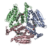
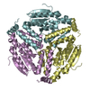
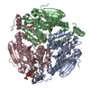
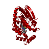
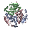
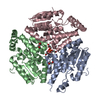
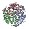
| Title | Crystal structure of M. tuberculosis EchA6 bound to C20-CoA | ||||||
 Components Components | enoyl-CoA hydratase echA6 | ||||||
 Keywords Keywords | LIPID BINDING PROTEIN /  enoyl-CoA hydratase-like / lyase enoyl-CoA hydratase-like / lyase | ||||||
| Function / homology |  Function and homology informationenoyl-CoA hydratase / enoyl-CoA hydratase activity / fatty acid beta-oxidation / peptidoglycan-based cell wall / plasma membrane Function and homology informationenoyl-CoA hydratase / enoyl-CoA hydratase activity / fatty acid beta-oxidation / peptidoglycan-based cell wall / plasma membraneSimilarity search - Function | ||||||
| Biological species |   Mycobacterium tuberculosis (bacteria) Mycobacterium tuberculosis (bacteria) | ||||||
| Method | X-RAY DIFFRACTION / SYNCHROTRON / MOLECULAR REPLACEMENT / Resolution: 2.439 Å | ||||||
 Authors Authors | Cox, J.A.G. / Besra, G.S. / Futterer, K. | ||||||
 Citation Citation | Journal: Nat Microbiol / Year: 2016 Title: THPP target assignment reveals EchA6 as an essential fatty acid shuttle in mycobacteria. Authors: Cox, J.A. / Abrahams, K.A. / Alemparte, C. / Ghidelli-Disse, S. / Rullas, J. / Angulo-Barturen, I. / Singh, A. / Gurcha, S.S. / Nataraj, V. / Bethell, S. / Remuinan, M.J. / Encinas, L. / ...Authors: Cox, J.A. / Abrahams, K.A. / Alemparte, C. / Ghidelli-Disse, S. / Rullas, J. / Angulo-Barturen, I. / Singh, A. / Gurcha, S.S. / Nataraj, V. / Bethell, S. / Remuinan, M.J. / Encinas, L. / Jervis, P.J. / Cammack, N.C. / Bhatt, A. / Kruse, U. / Bantscheff, M. / Futterer, K. / Barros, D. / Ballell, L. / Drewes, G. / Besra, G.S. | ||||||
| History |
|
- Structure visualization
| Structure viewer | Molecule: MolmilJmol/JSmol |
|---|
- Downloads & links
-Download
| PDBx/mmCIF format | 5dtw.cif.gz | 535.4 KB | Display | PDBx/mmCIF format |
|---|---|---|---|---|
| PDB format | pdb5dtw.ent.gz | 442.8 KB | Display | PDB format |
| PDBx/mmJSON format | 5dtw.json.gz | Tree view | PDBx/mmJSON format | |
| Others |  Other downloads Other downloads |
-Validation report
| Arichive directory | https://data.pdbj.org/pub/pdb/validation_reports/dt/5dtwftp://data.pdbj.org/pub/pdb/validation_reports/dt/5dtw | HTTPS FTP |
|---|
-Related structure data
| Related structure data |  5dtpC  5du4C 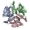 5du6C 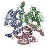 5du8C  5ducC  5dufC 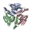 3he2S C: citing same article ( S: Starting model for refinement |
|---|---|
| Similar structure data |


-Links
 PDBj
PDBj- Assembly
| Deposited unit | 
| ||||||||
|---|---|---|---|---|---|---|---|---|---|
| 1 | 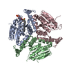
| ||||||||
| 2 | 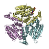
| ||||||||
| Unit cell |
|
-Components
| #1: Protein | Mass: 28230.186 Da / Num. of mol.: 6 Source method: isolated from a genetically manipulated source Source: (gene. exp.) Mycobacterium tuberculosis (strain ATCC 25618 / H37Rv) (bacteria)Strain: ATCC 25618 / H37Rv / Gene: echA6, Rv0905, MTCY31.33 / Production host: Escherichia coli BL21(DE3) (bacteria) / References: UniProt: P9WNP1, enoyl-CoA hydratase#2: Chemical |   Mass: 1062.049 Da / Num. of mol.: 2 / Source method: obtained synthetically / Formula: C41H74N7O17P3S Mass: 1062.049 Da / Num. of mol.: 2 / Source method: obtained synthetically / Formula: C41H74N7O17P3S#3: Water | ChemComp-HOH / | Water Mass: 18.015 Da / Num. of mol.: 121 / Source method: isolated from a natural source / Formula: H2O Mass: 18.015 Da / Num. of mol.: 121 / Source method: isolated from a natural source / Formula: H2O |
|---|
-Experimental details
-Experiment
| Experiment | Method: X-RAY DIFFRACTION |
|---|
- Sample preparation
| Crystal | Density Matthews: 2.8 Å3/Da / Density % sol: 56.01 % |
|---|---|
| Crystal grow | Temperature: 291 K / Method: vapor diffusion, sitting drop Details: 0.17 M ammonium sulfate, 25.5 % w/v PEG 4000, 15 % v/v glycerol |
-Data collection
| Diffraction | Mean temperature: 100 K |
|---|---|
| Diffraction source | Source: SYNCHROTRON / Site: Diamond  / Beamline: I03 / Wavelength: 0.9763 Å / Beamline: I03 / Wavelength: 0.9763 Å |
| Detector | Type: DECTRIS PILATUS 6M / Detector: PIXEL / Date: Feb 2, 2013 |
| Radiation | Protocol: SINGLE WAVELENGTH / Monochromatic (M) / Laue (L): M / Scattering type: x-ray |
| Radiation wavelength | Wavelength: 0.9763 Å / Relative weight: 1 |
| Reflection | Resolution: 2.4→108.9 Å / Num. all: 246225 / Num. obs: 68420 / % possible obs: 99.4 % / Observed criterion σ(F): 0 / Observed criterion σ(I): 0 / Redundancy: 3.6 % / Rmerge(I) obs: 0.084 / Rsym value: 0.084 / Net I/av σ(I): 9.6 / Net I/σ(I): 9.6 |
| Reflection shell | Resolution: 2.4→2.46 Å / Redundancy: 3.3 % / Rmerge(I) obs: 0.499 / Mean I/σ(I) obs: 2.1 / % possible all: 99.5 |
- Processing
| Software |
| |||||||||||||||||||||||||||||||||||||||||||||||||||||||||||||||||||||||||||||||||||||||||||||||||||||||||||||||||||||||||||||||||||||||||||||||||||||||||||||||||||||||||||||||
|---|---|---|---|---|---|---|---|---|---|---|---|---|---|---|---|---|---|---|---|---|---|---|---|---|---|---|---|---|---|---|---|---|---|---|---|---|---|---|---|---|---|---|---|---|---|---|---|---|---|---|---|---|---|---|---|---|---|---|---|---|---|---|---|---|---|---|---|---|---|---|---|---|---|---|---|---|---|---|---|---|---|---|---|---|---|---|---|---|---|---|---|---|---|---|---|---|---|---|---|---|---|---|---|---|---|---|---|---|---|---|---|---|---|---|---|---|---|---|---|---|---|---|---|---|---|---|---|---|---|---|---|---|---|---|---|---|---|---|---|---|---|---|---|---|---|---|---|---|---|---|---|---|---|---|---|---|---|---|---|---|---|---|---|---|---|---|---|---|---|---|---|---|---|---|---|---|
| Refinement | Method to determine structure: MOLECULAR REPLACEMENT Starting model: 3HE2 Resolution: 2.439→42.848 Å / SU ML: 0.42 / Cross valid method: FREE R-VALUE / σ(F): 1.18 / Phase error: 27.22 / Stereochemistry target values: ML
| |||||||||||||||||||||||||||||||||||||||||||||||||||||||||||||||||||||||||||||||||||||||||||||||||||||||||||||||||||||||||||||||||||||||||||||||||||||||||||||||||||||||||||||||
| Solvent computation | Shrinkage radii: 1.11 Å / VDW probe radii: 1.3 Å / Solvent model: FLAT BULK SOLVENT MODEL / Bsol: 8.26 Å2 / ksol: 0.322 e/Å3 | |||||||||||||||||||||||||||||||||||||||||||||||||||||||||||||||||||||||||||||||||||||||||||||||||||||||||||||||||||||||||||||||||||||||||||||||||||||||||||||||||||||||||||||||
| Displacement parameters |
| |||||||||||||||||||||||||||||||||||||||||||||||||||||||||||||||||||||||||||||||||||||||||||||||||||||||||||||||||||||||||||||||||||||||||||||||||||||||||||||||||||||||||||||||
| Refinement step | Cycle: LAST / Resolution: 2.439→42.848 Å
| |||||||||||||||||||||||||||||||||||||||||||||||||||||||||||||||||||||||||||||||||||||||||||||||||||||||||||||||||||||||||||||||||||||||||||||||||||||||||||||||||||||||||||||||
| Refine LS restraints |
| |||||||||||||||||||||||||||||||||||||||||||||||||||||||||||||||||||||||||||||||||||||||||||||||||||||||||||||||||||||||||||||||||||||||||||||||||||||||||||||||||||||||||||||||
| LS refinement shell |
| |||||||||||||||||||||||||||||||||||||||||||||||||||||||||||||||||||||||||||||||||||||||||||||||||||||||||||||||||||||||||||||||||||||||||||||||||||||||||||||||||||||||||||||||
| Refinement TLS params. | Method: refined / Refine-ID: X-RAY DIFFRACTION
| |||||||||||||||||||||||||||||||||||||||||||||||||||||||||||||||||||||||||||||||||||||||||||||||||||||||||||||||||||||||||||||||||||||||||||||||||||||||||||||||||||||||||||||||
| Refinement TLS group |
|
