 Movie
Movie Controller


+ Open data

- Basic information
| Entry | Database: PDB / ID: 3vcm | ||||||
|---|---|---|---|---|---|---|---|
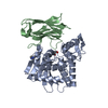
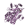
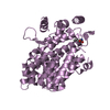
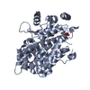
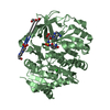
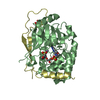
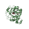
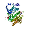
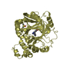
| Title | Crystal structure of human prorenin | ||||||
 Components Components | (prorenin ) x 2 ) x 2 | ||||||
 Keywords Keywords | HYDROLASE / aspartic proteases / prorenin receptor | ||||||
| Function / homology |  Function and homology informationrenin / juxtaglomerular apparatus development / mesonephros development / response to cGMP / renin-angiotensin regulation of aldosterone production / drinking behavior / regulation of MAPK cascade / response to immobilization stress / angiotensin maturation / amyloid-beta metabolic process ...renin / juxtaglomerular apparatus development / mesonephros development / response to cGMP / renin-angiotensin regulation of aldosterone production / drinking behavior / regulation of MAPK cascade / response to immobilization stress / angiotensin maturation / amyloid-beta metabolic process / Metabolism of Angiotensinogen to Angiotensins / cell maturation / response to cAMP / insulin-like growth factor receptor binding / hormone-mediated signaling pathway / kidney development / regulation of blood pressure / male gonad development / cellular response to xenobiotic stimulus / apical part of cell / peptidase activity / response to lipopolysaccharide / aspartic-type endopeptidase activity / signaling receptor binding / proteolysis / extracellular space / extracellular region / plasma membrane Function and homology informationrenin / juxtaglomerular apparatus development / mesonephros development / response to cGMP / renin-angiotensin regulation of aldosterone production / drinking behavior / regulation of MAPK cascade / response to immobilization stress / angiotensin maturation / amyloid-beta metabolic process ...renin / juxtaglomerular apparatus development / mesonephros development / response to cGMP / renin-angiotensin regulation of aldosterone production / drinking behavior / regulation of MAPK cascade / response to immobilization stress / angiotensin maturation / amyloid-beta metabolic process / Metabolism of Angiotensinogen to Angiotensins / cell maturation / response to cAMP / insulin-like growth factor receptor binding / hormone-mediated signaling pathway / kidney development / regulation of blood pressure / male gonad development / cellular response to xenobiotic stimulus / apical part of cell / peptidase activity / response to lipopolysaccharide / aspartic-type endopeptidase activity / signaling receptor binding / proteolysis / extracellular space / extracellular region / plasma membraneSimilarity search - Function | ||||||
| Biological species |  Homo sapiens (human) Homo sapiens (human) | ||||||
| Method | X-RAY DIFFRACTION / SYNCHROTRON / MOLECULAR REPLACEMENT / Resolution: 2.93 Å | ||||||
 Authors Authors | Morales, R. / Watier, Y. / Bocskei, Z. | ||||||
 Citation Citation | Journal: J.Mol.Biol. / Year: 2012 Title: Human Prorenin Structure Sheds Light on a Novel Mechanism of Its Autoinhibition and on Its Non-Proteolytic Activation by the (Pro)renin Receptor. Authors: Morales, R. / Watier, Y. / Bocskei, Z. | ||||||
| History |
|
- Structure visualization
| Structure viewer | Molecule: MolmilJmol/JSmol |
|---|
- Downloads & links
-Download
| PDBx/mmCIF format | 3vcm.cif.gz | 289.8 KB | Display | PDBx/mmCIF format |
|---|---|---|---|---|
| PDB format | pdb3vcm.ent.gz | 236.3 KB | Display | PDB format |
| PDBx/mmJSON format | 3vcm.json.gz | Tree view | PDBx/mmJSON format | |
| Others |  Other downloads Other downloads |
-Validation report
| Arichive directory | https://data.pdbj.org/pub/pdb/validation_reports/vc/3vcmftp://data.pdbj.org/pub/pdb/validation_reports/vc/3vcm | HTTPS FTP |
|---|
-Related structure data
| Similar structure data |
|---|

-Links
 PDBj
PDBj


- Assembly
| Deposited unit | 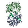
| ||||||||
|---|---|---|---|---|---|---|---|---|---|
| 1 | 
| ||||||||
| 2 | 
| ||||||||
| Unit cell |
|
-Components
| #1: Protein | / Angiotensinogenase Mass: 36721.508 Da / Num. of mol.: 2 / Fragment: renin (UNP residues 67-406) Source method: isolated from a genetically manipulated source Source: (gene. exp.) Homo sapiens (human) / Gene: REN / Cell line (production host): HEK / Production host: Homo sapiens (human) / References: UniProt: P00797, renin#2: Protein/peptide | / AngiotensinogenaseMass: 5115.053 Da / Num. of mol.: 2 / Fragment: activation peptide (UNP residues 24-66) Source method: isolated from a genetically manipulated source Source: (gene. exp.) Homo sapiens (human) / Gene: REN / Cell line (production host): HEK / Production host: Homo sapiens (human) / References: UniProt: P00797, renin#3: Sugar | ChemComp-NAG / | N-Acetylglucosamine  Type: D-saccharide, beta linking / Mass: 221.208 Da / Num. of mol.: 1 Type: D-saccharide, beta linking / Mass: 221.208 Da / Num. of mol.: 1Source method: isolated from a genetically manipulated source Formula: C8H15NO6 #4: Water | ChemComp-HOH / | Water Mass: 18.015 Da / Num. of mol.: 97 / Source method: isolated from a natural source / Formula: H2O Mass: 18.015 Da / Num. of mol.: 97 / Source method: isolated from a natural source / Formula: H2O |
|---|
-Experimental details
-Experiment
| Experiment | Method: X-RAY DIFFRACTION / Number of used crystals: 1 |
|---|
- Sample preparation
| Crystal | Density Matthews: 3.86 Å3/Da / Density % sol: 68.15 % |
|---|---|
| Crystal grow | Temperature: 298 K / Method: vapor diffusion, sitting drop / pH: 7 Details: 1.5 M sodium malonate, 0.1 M Bis-Tris-propane, pH 7.0, VAPOR DIFFUSION, SITTING DROP, temperature 298K |
-Data collection
| Diffraction | Mean temperature: 173 K |
|---|---|
| Diffraction source | Source: SYNCHROTRON / Site: ESRF  / Beamline: ID23-2 / Beamline: ID23-2 |
| Detector | Type: MARMOSAIC 225 mm CCD / Detector: CCD |
| Radiation | Monochromator: Si(111) / Protocol: SINGLE WAVELENGTH / Monochromatic (M) / Laue (L): M / Scattering type: x-ray |
| Radiation wavelength | Relative weight: 1 |
| Reflection | Resolution: 2.93→50 Å / Num. obs: 29148 / % possible obs: 100 % / Redundancy: 6 % / Biso Wilson estimate: 61.96 Å2 / Rmerge(I) obs: 0.129 / Net I/σ(I): 12.1 |
| Reflection shell | Resolution: 2.93→3.09 Å / Redundancy: 6.2 % / Rmerge(I) obs: 0.513 / Mean I/σ(I) obs: 3.8 / Num. unique all: 4176 / % possible all: 100 |
- Processing
| Software |
| |||||||||||||||||||||||||||||||||||||||||||||||||||||||||||||||||||||||||||||||||||||||||||||||||||||||||||||||||||||||||||||
|---|---|---|---|---|---|---|---|---|---|---|---|---|---|---|---|---|---|---|---|---|---|---|---|---|---|---|---|---|---|---|---|---|---|---|---|---|---|---|---|---|---|---|---|---|---|---|---|---|---|---|---|---|---|---|---|---|---|---|---|---|---|---|---|---|---|---|---|---|---|---|---|---|---|---|---|---|---|---|---|---|---|---|---|---|---|---|---|---|---|---|---|---|---|---|---|---|---|---|---|---|---|---|---|---|---|---|---|---|---|---|---|---|---|---|---|---|---|---|---|---|---|---|---|---|---|---|
| Refinement | Method to determine structure: MOLECULAR REPLACEMENT / Resolution: 2.93→36.53 Å / Cor.coef. Fo:Fc: 0.8729 / Cor.coef. Fo:Fc free: 0.8289 / SU R Cruickshank DPI: 0.599 / Cross valid method: THROUGHOUT / σ(F): 0 / SU R Blow DPI: 0.634 / SU Rfree Blow DPI: 0.321 / SU Rfree Cruickshank DPI: 0.322
| |||||||||||||||||||||||||||||||||||||||||||||||||||||||||||||||||||||||||||||||||||||||||||||||||||||||||||||||||||||||||||||
| Displacement parameters | Biso mean: 48.32 Å2
| |||||||||||||||||||||||||||||||||||||||||||||||||||||||||||||||||||||||||||||||||||||||||||||||||||||||||||||||||||||||||||||
| Refine analyze | Luzzati coordinate error obs: 0.405 Å | |||||||||||||||||||||||||||||||||||||||||||||||||||||||||||||||||||||||||||||||||||||||||||||||||||||||||||||||||||||||||||||
| Refinement step | Cycle: LAST / Resolution: 2.93→36.53 Å
| |||||||||||||||||||||||||||||||||||||||||||||||||||||||||||||||||||||||||||||||||||||||||||||||||||||||||||||||||||||||||||||
| Refine LS restraints |
| |||||||||||||||||||||||||||||||||||||||||||||||||||||||||||||||||||||||||||||||||||||||||||||||||||||||||||||||||||||||||||||
| LS refinement shell | Resolution: 2.93→3.03 Å / Total num. of bins used: 15
| |||||||||||||||||||||||||||||||||||||||||||||||||||||||||||||||||||||||||||||||||||||||||||||||||||||||||||||||||||||||||||||
| Refinement TLS params. | Method: refined / Refine-ID: X-RAY DIFFRACTION
| |||||||||||||||||||||||||||||||||||||||||||||||||||||||||||||||||||||||||||||||||||||||||||||||||||||||||||||||||||||||||||||
| Refinement TLS group |
|
