 Movie
Movie Controller


+ Open data

- Basic information
| Entry | Database: PDB / ID: 5xvf | ||||||
|---|---|---|---|---|---|---|---|
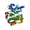
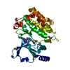
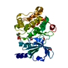
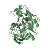
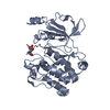
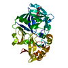
| Title | Crystal Structure of PAK4 in complex with inhibitor CZH062 | ||||||
 Components Components | Serine/threonine-protein kinase PAK 4 | ||||||
 Keywords Keywords |  TRANSFERASE / ATP binding pocket TRANSFERASE / ATP binding pocket | ||||||
| Function / homology |  Function and homology information Function and homology informationdendritic spine development / cadherin binding involved in cell-cell adhesion / Activation of RAC1 / RHOV GTPase cycle / RHOJ GTPase cycle / RHOQ GTPase cycle / regulation of MAPK cascade / RHOH GTPase cycle / CDC42 GTPase cycle / RHOU GTPase cycle ...dendritic spine development / cadherin binding involved in cell-cell adhesion / Activation of RAC1 / RHOV GTPase cycle / RHOJ GTPase cycle / RHOQ GTPase cycle / regulation of MAPK cascade / RHOH GTPase cycle / CDC42 GTPase cycle / RHOU GTPase cycle / cellular response to organic cyclic compound / RHOG GTPase cycle / RAC2 GTPase cycle / RAC3 GTPase cycle / negative regulation of endothelial cell apoptotic process / cytoskeleton organization / RAC1 GTPase cycle / regulation of cell growth / adherens junction / positive regulation of angiogenesis / cell migration / non-specific serine/threonine protein kinase / protein kinase activity / intracellular signal transduction / cell cycle / phosphorylation / protein serine kinase activity / focal adhesion / protein serine/threonine kinase activity / apoptotic process / Golgi apparatus / signal transduction / ATP binding / cytosol / cytoplasmSimilarity search - Function | ||||||
| Biological species |  Homo sapiens (human) Homo sapiens (human) | ||||||
| Method | X-RAY DIFFRACTION / SYNCHROTRON / MOLECULAR REPLACEMENT / Resolution: 2.655 Å | ||||||
 Authors Authors | Zhao, F. / Li, H. | ||||||
 Citation Citation | Journal: J. Med. Chem. / Year: 2018 Title: Structure-Based Design of 6-Chloro-4-aminoquinazoline-2-carboxamide Derivatives as Potent and Selective p21-Activated Kinase 4 (PAK4) Inhibitors. Authors: Hao, C. / Zhao, F. / Song, H. / Guo, J. / Li, X. / Jiang, X. / Huan, R. / Song, S. / Zhang, Q. / Wang, R. / Wang, K. / Pang, Y. / Liu, T. / Lu, T. / Huang, W. / Wang, J. / Lin, B. / He, Z. / ...Authors: Hao, C. / Zhao, F. / Song, H. / Guo, J. / Li, X. / Jiang, X. / Huan, R. / Song, S. / Zhang, Q. / Wang, R. / Wang, K. / Pang, Y. / Liu, T. / Lu, T. / Huang, W. / Wang, J. / Lin, B. / He, Z. / Li, H. / Li, F. / Zhao, D. / Cheng, M. | ||||||
| History |
|
- Structure visualization
| Structure viewer | Molecule: MolmilJmol/JSmol |
|---|
- Downloads & links
-Download
| PDBx/mmCIF format | 5xvf.cif.gz | 132.5 KB | Display | PDBx/mmCIF format |
|---|---|---|---|---|
| PDB format | pdb5xvf.ent.gz | 103.6 KB | Display | PDB format |
| PDBx/mmJSON format | 5xvf.json.gz | Tree view | PDBx/mmJSON format | |
| Others |  Other downloads Other downloads |
-Validation report
| Arichive directory | https://data.pdbj.org/pub/pdb/validation_reports/xv/5xvfftp://data.pdbj.org/pub/pdb/validation_reports/xv/5xvf | HTTPS FTP |
|---|
-Related structure data
| Related structure data |  5xvaC  5xvgC  2j0iS S: Starting model for refinement C: citing same article ( |
|---|---|
| Similar structure data |


-Links
 PDBj
PDBj


- Assembly
| Deposited unit | 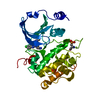
| ||||||||
|---|---|---|---|---|---|---|---|---|---|
| 1 |
| ||||||||
| Unit cell |
|
-Components
| #1: Protein | Mass: 32682.955 Da / Num. of mol.: 1 / Fragment: PAK4 Kinase Domain (UNP RESIDUES 300-588) Source method: isolated from a genetically manipulated source Source: (gene. exp.) Homo sapiens (human) / Gene: PAK4, KIAA1142 / Plasmid: pSUMOH10 / Production host:  Escherichia coli (E. coli) / Strain (production host): BL21(DE3) Escherichia coli (E. coli) / Strain (production host): BL21(DE3)References: UniProt: O96013, non-specific serine/threonine protein kinase |
|---|---|
| #2: Chemical | ChemComp-8FR /   Mass: 357.841 Da / Num. of mol.: 1 / Source method: obtained synthetically / Formula: C17H20ClN7 Mass: 357.841 Da / Num. of mol.: 1 / Source method: obtained synthetically / Formula: C17H20ClN7 |
| #3: Water | ChemComp-HOH / Water Mass: 18.015 Da / Num. of mol.: 7 / Source method: isolated from a natural source / Formula: H2O Mass: 18.015 Da / Num. of mol.: 7 / Source method: isolated from a natural source / Formula: H2O |
-Experimental details
-Experiment
| Experiment | Method: X-RAY DIFFRACTION / Number of used crystals: 1 |
|---|
- Sample preparation
| Crystal | Density Matthews: 2.96 Å3/Da / Density % sol: 58.47 % / Mosaicity: 0.632 ° / Mosaicity esd: 0.008 ° |
|---|---|
| Crystal grow | Temperature: 291 K / Method: vapor diffusion / pH: 7.5 / Details: 0.1 M HEPES pH 7.5, 25% PEG3350. |
-Data collection
| Diffraction | Mean temperature: 100 K | |||||||||||||||||||||||||||||||||||||||||||||||||||||||||||||||||||||||||||||||||||||||||||||||||||||||||||||||||||||||||||||||||||||||||||||||||||||||||||||||||||||||||||||||||||||||||||||
|---|---|---|---|---|---|---|---|---|---|---|---|---|---|---|---|---|---|---|---|---|---|---|---|---|---|---|---|---|---|---|---|---|---|---|---|---|---|---|---|---|---|---|---|---|---|---|---|---|---|---|---|---|---|---|---|---|---|---|---|---|---|---|---|---|---|---|---|---|---|---|---|---|---|---|---|---|---|---|---|---|---|---|---|---|---|---|---|---|---|---|---|---|---|---|---|---|---|---|---|---|---|---|---|---|---|---|---|---|---|---|---|---|---|---|---|---|---|---|---|---|---|---|---|---|---|---|---|---|---|---|---|---|---|---|---|---|---|---|---|---|---|---|---|---|---|---|---|---|---|---|---|---|---|---|---|---|---|---|---|---|---|---|---|---|---|---|---|---|---|---|---|---|---|---|---|---|---|---|---|---|---|---|---|---|---|---|---|---|---|---|
| Diffraction source | Source: SYNCHROTRON / Site: SSRF  / Beamline: BL17U1 / Wavelength: 1.2818 Å / Beamline: BL17U1 / Wavelength: 1.2818 Å | |||||||||||||||||||||||||||||||||||||||||||||||||||||||||||||||||||||||||||||||||||||||||||||||||||||||||||||||||||||||||||||||||||||||||||||||||||||||||||||||||||||||||||||||||||||||||||||
| Detector | Type: ADSC QUANTUM 315r / Detector: CCD / Date: Jan 9, 2016 / Details: mirrors | |||||||||||||||||||||||||||||||||||||||||||||||||||||||||||||||||||||||||||||||||||||||||||||||||||||||||||||||||||||||||||||||||||||||||||||||||||||||||||||||||||||||||||||||||||||||||||||
| Radiation | Monochromator: double crystal, Si(111) / Protocol: SINGLE WAVELENGTH / Monochromatic (M) / Laue (L): M / Scattering type: x-ray | |||||||||||||||||||||||||||||||||||||||||||||||||||||||||||||||||||||||||||||||||||||||||||||||||||||||||||||||||||||||||||||||||||||||||||||||||||||||||||||||||||||||||||||||||||||||||||||
| Radiation wavelength | Wavelength: 1.2818 Å / Relative weight: 1 | |||||||||||||||||||||||||||||||||||||||||||||||||||||||||||||||||||||||||||||||||||||||||||||||||||||||||||||||||||||||||||||||||||||||||||||||||||||||||||||||||||||||||||||||||||||||||||||
| Reflection | Resolution: 2.65→50 Å / Num. obs: 12074 / % possible obs: 99.9 % / Redundancy: 11 % / Biso Wilson estimate: 65.77 Å2 / Rmerge(I) obs: 0.088 / Rpim(I) all: 0.028 / Rrim(I) all: 0.092 / Χ2: 1.968 / Net I/σ(I): 10.6 / Num. measured all: 132467 | |||||||||||||||||||||||||||||||||||||||||||||||||||||||||||||||||||||||||||||||||||||||||||||||||||||||||||||||||||||||||||||||||||||||||||||||||||||||||||||||||||||||||||||||||||||||||||||
| Reflection shell | Diffraction-ID: 1
|
- Processing
| Software |
| |||||||||||||||||||||||||||||||||||||||||||||||||||||||||||||||||||||||||||||||||||||||||||||||||||||||||||||||||||||||||||||
|---|---|---|---|---|---|---|---|---|---|---|---|---|---|---|---|---|---|---|---|---|---|---|---|---|---|---|---|---|---|---|---|---|---|---|---|---|---|---|---|---|---|---|---|---|---|---|---|---|---|---|---|---|---|---|---|---|---|---|---|---|---|---|---|---|---|---|---|---|---|---|---|---|---|---|---|---|---|---|---|---|---|---|---|---|---|---|---|---|---|---|---|---|---|---|---|---|---|---|---|---|---|---|---|---|---|---|---|---|---|---|---|---|---|---|---|---|---|---|---|---|---|---|---|---|---|---|
| Refinement | Method to determine structure: MOLECULAR REPLACEMENT Starting model: 2j0i Resolution: 2.655→37.193 Å / SU ML: 0.31 / Cross valid method: THROUGHOUT / σ(F): 1.36 / Phase error: 27.46 / Stereochemistry target values: ML
| |||||||||||||||||||||||||||||||||||||||||||||||||||||||||||||||||||||||||||||||||||||||||||||||||||||||||||||||||||||||||||||
| Solvent computation | Shrinkage radii: 0.9 Å / VDW probe radii: 1.11 Å / Solvent model: FLAT BULK SOLVENT MODEL | |||||||||||||||||||||||||||||||||||||||||||||||||||||||||||||||||||||||||||||||||||||||||||||||||||||||||||||||||||||||||||||
| Displacement parameters | Biso max: 146.61 Å2 / Biso mean: 74.48 Å2 / Biso min: 30 Å2 | |||||||||||||||||||||||||||||||||||||||||||||||||||||||||||||||||||||||||||||||||||||||||||||||||||||||||||||||||||||||||||||
| Refinement step | Cycle: final / Resolution: 2.655→37.193 Å
| |||||||||||||||||||||||||||||||||||||||||||||||||||||||||||||||||||||||||||||||||||||||||||||||||||||||||||||||||||||||||||||
| Refine LS restraints |
| |||||||||||||||||||||||||||||||||||||||||||||||||||||||||||||||||||||||||||||||||||||||||||||||||||||||||||||||||||||||||||||
| LS refinement shell | Refine-ID: X-RAY DIFFRACTION / Rfactor Rfree error: 0 / Total num. of bins used: 4
| |||||||||||||||||||||||||||||||||||||||||||||||||||||||||||||||||||||||||||||||||||||||||||||||||||||||||||||||||||||||||||||
| Refinement TLS params. | Method: refined / Refine-ID: X-RAY DIFFRACTION
| |||||||||||||||||||||||||||||||||||||||||||||||||||||||||||||||||||||||||||||||||||||||||||||||||||||||||||||||||||||||||||||
| Refinement TLS group |
|
