 Movie
Movie Controller


+ Open data

- Basic information
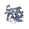
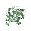
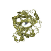
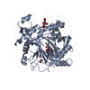
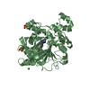
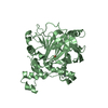
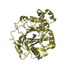
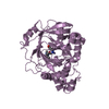
| Entry | Database: PDB / ID: 6g5x | ||||||
|---|---|---|---|---|---|---|---|
| Title | Crystal Structure of KDM4A with compound YP-02-145 | ||||||
 Components Components | Lysine-specific demethylase 4A | ||||||
 Keywords Keywords |  OXIDOREDUCTASE / KDM4A / ligand binding / drug design / inhibitor design / cancer / epigenetics OXIDOREDUCTASE / KDM4A / ligand binding / drug design / inhibitor design / cancer / epigenetics | ||||||
| Function / homology |  Function and homology information Function and homology information[histone H3]-trimethyl-L-lysine36 demethylase / histone H3K36me2/H3K36me3 demethylase activity / apoptotic chromosome condensation / histone H3K36 demethylase activity / cardiac muscle hypertrophy in response to stress / histone H3K9me2/H3K9me3 demethylase activity / [histone H3]-trimethyl-L-lysine9 demethylase / histone H3K9 demethylase activity / negative regulation of astrocyte differentiation / histone demethylase activity ...[histone H3]-trimethyl-L-lysine36 demethylase / histone H3K36me2/H3K36me3 demethylase activity / apoptotic chromosome condensation / histone H3K36 demethylase activity / cardiac muscle hypertrophy in response to stress / histone H3K9me2/H3K9me3 demethylase activity / [histone H3]-trimethyl-L-lysine9 demethylase / histone H3K9 demethylase activity / negative regulation of astrocyte differentiation / histone demethylase activity / pericentric heterochromatin / NR1H3 & NR1H2 regulate gene expression linked to cholesterol transport and efflux / methylated histone binding / positive regulation of neuron differentiation / response to nutrient levels / negative regulation of autophagy / HDMs demethylate histones / fibrillar center / Recruitment and ATM-mediated phosphorylation of repair and signaling proteins at DNA double strand breaks / regulation of gene expression / chromatin remodeling / negative regulation of gene expression / negative regulation of DNA-templated transcription / ubiquitin protein ligase binding / chromatin / positive regulation of gene expression / zinc ion binding / nucleoplasm / nucleus / cytosolSimilarity search - Function | ||||||
| Biological species |  Homo sapiens (human) Homo sapiens (human) | ||||||
| Method | X-RAY DIFFRACTION / SYNCHROTRON / MOLECULAR REPLACEMENT / Resolution: 1.78 Å | ||||||
 Authors Authors | Malecki, P.H. / Carter, D.M. / Gohlke, U. / Specker, E. / Nazare, M. / Weiss, M.S. / Heinemann, U. | ||||||
 Citation Citation | Journal: J.Med.Chem. / Year: 2021 Title: Enhanced Properties of a Benzimidazole Benzylpyrazole Lysine Demethylase Inhibitor: Mechanism-of-Action, Binding Site Analysis, and Activity in Cellular Models of Prostate Cancer. Authors: Carter, D.M. / Specker, E. / Malecki, P.H. / Przygodda, J. / Dudaniec, K. / Weiss, M.S. / Heinemann, U. / Nazare, M. / Gohlke, U. | ||||||
| History |
|
- Structure visualization
| Structure viewer | Molecule: MolmilJmol/JSmol |
|---|
- Downloads & links
-Download
| PDBx/mmCIF format | 6g5x.cif.gz | 327.8 KB | Display | PDBx/mmCIF format |
|---|---|---|---|---|
| PDB format | pdb6g5x.ent.gz | 266.1 KB | Display | PDB format |
| PDBx/mmJSON format | 6g5x.json.gz | Tree view | PDBx/mmJSON format | |
| Others |  Other downloads Other downloads |
-Validation report
| Arichive directory | https://data.pdbj.org/pub/pdb/validation_reports/g5/6g5xftp://data.pdbj.org/pub/pdb/validation_reports/g5/6g5x | HTTPS FTP |
|---|
-Related structure data
| Related structure data |  6g5wC 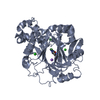 3pdqS S: Starting model for refinement C: citing same article ( |
|---|---|
| Similar structure data |


-Links
 PDBj
PDBj


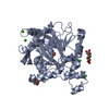
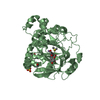
- Assembly
| Deposited unit | 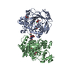
| ||||||||
|---|---|---|---|---|---|---|---|---|---|
| 1 | 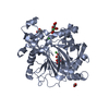
| ||||||||
| 2 | 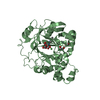
| ||||||||
| Unit cell |
|
-Components
-Protein , 1 types, 2 molecules AB
| #1: Protein | Mass: 41767.543 Da / Num. of mol.: 2 / Fragment: JMJD2A Source method: isolated from a genetically manipulated source Source: (gene. exp.) Homo sapiens (human) / Gene: KDM4A, JHDM3A, JMJD2, JMJD2A, KIAA0677 / Production host:  Escherichia coli (E. coli) / Strain (production host): BL21(DE3) Escherichia coli (E. coli) / Strain (production host): BL21(DE3)References: UniProt: O75164, Oxidoreductases; Acting on paired donors, with incorporation or reduction of molecular oxygen; With 2-oxoglutarate as one donor, and incorporation of one atom of oxygen into each donor |
|---|
-Non-polymers , 7 types, 566 molecules 


| #2: Chemical |  Mass: 65.409 Da / Num. of mol.: 2 / Source method: obtained synthetically / Formula: Zn Mass: 65.409 Da / Num. of mol.: 2 / Source method: obtained synthetically / Formula: Zn#3: Chemical | ChemComp-NI / Nickel Mass: 58.693 Da / Num. of mol.: 4 / Source method: obtained synthetically / Formula: Ni Mass: 58.693 Da / Num. of mol.: 4 / Source method: obtained synthetically / Formula: Ni#4: Chemical | ChemComp-EDO / Ethylene glycol Mass: 62.068 Da / Num. of mol.: 4 / Source method: obtained synthetically / Formula: C2H6O2 Mass: 62.068 Da / Num. of mol.: 4 / Source method: obtained synthetically / Formula: C2H6O2#5: Chemical | Citric acid Mass: 192.124 Da / Num. of mol.: 3 / Source method: obtained synthetically / Formula: C6H8O7 Mass: 192.124 Da / Num. of mol.: 3 / Source method: obtained synthetically / Formula: C6H8O7#6: Chemical | ChemComp-GOL / | Glycerol Mass: 92.094 Da / Num. of mol.: 1 / Source method: obtained synthetically / Formula: C3H8O3 Mass: 92.094 Da / Num. of mol.: 1 / Source method: obtained synthetically / Formula: C3H8O3#7: Chemical | ChemComp-MY7 / |  Mass: 334.329 Da / Num. of mol.: 1 / Source method: obtained synthetically / Formula: C18H14N4O3 Mass: 334.329 Da / Num. of mol.: 1 / Source method: obtained synthetically / Formula: C18H14N4O3#8: Water | ChemComp-HOH / | WaterMass: 18.015 Da / Num. of mol.: 551 / Source method: isolated from a natural source / Formula: H2O |
|---|
-Experimental details
-Experiment
| Experiment | Method: X-RAY DIFFRACTION / Number of used crystals: 1 |
|---|
- Sample preparation
| Crystal | Density Matthews: 2.51 Å3/Da / Density % sol: 50.98 % |
|---|---|
| Crystal grow | Temperature: 277 K / Method: vapor diffusion / pH: 5.5 / Details: 100mM Citrate, 10mM NiCl2, 20% PEG 3350 |
-Data collection
| Diffraction | Mean temperature: 100 K | ||||||||||||||||||||||||||||||||||||||||||||||||||||||||||||||||||||||||||||||||
|---|---|---|---|---|---|---|---|---|---|---|---|---|---|---|---|---|---|---|---|---|---|---|---|---|---|---|---|---|---|---|---|---|---|---|---|---|---|---|---|---|---|---|---|---|---|---|---|---|---|---|---|---|---|---|---|---|---|---|---|---|---|---|---|---|---|---|---|---|---|---|---|---|---|---|---|---|---|---|---|---|---|
| Diffraction source | Source: SYNCHROTRON / Site: BESSY  / Beamline: 14.1 / Wavelength: 0.9184 Å / Beamline: 14.1 / Wavelength: 0.9184 Å | ||||||||||||||||||||||||||||||||||||||||||||||||||||||||||||||||||||||||||||||||
| Detector | Type: DECTRIS PILATUS 6M / Detector: PIXEL / Date: Sep 4, 2015 | ||||||||||||||||||||||||||||||||||||||||||||||||||||||||||||||||||||||||||||||||
| Radiation | Monochromator: DOUBLE CRYSTAL / Protocol: SINGLE WAVELENGTH / Monochromatic (M) / Laue (L): M / Scattering type: x-ray | ||||||||||||||||||||||||||||||||||||||||||||||||||||||||||||||||||||||||||||||||
| Radiation wavelength | Wavelength: 0.9184 Å / Relative weight: 1 | ||||||||||||||||||||||||||||||||||||||||||||||||||||||||||||||||||||||||||||||||
| Reflection | Resolution: 1.78→46.84 Å / Num. obs: 81224 / % possible obs: 99.7 % / Redundancy: 5.891 % / Biso Wilson estimate: 28.7 Å2 / CC1/2: 1 / Rmerge(I) obs: 0.051 / Rrim(I) all: 0.056 / Χ2: 1.002 / Net I/σ(I): 21.54 | ||||||||||||||||||||||||||||||||||||||||||||||||||||||||||||||||||||||||||||||||
| Reflection shell | Diffraction-ID: 1
|
- Processing
| Software |
| ||||||||||||||||||||||||||||||||||||||||||||||||||||||||||||||||||||||||||||||||||||||||||||||||||||||||||||||||
|---|---|---|---|---|---|---|---|---|---|---|---|---|---|---|---|---|---|---|---|---|---|---|---|---|---|---|---|---|---|---|---|---|---|---|---|---|---|---|---|---|---|---|---|---|---|---|---|---|---|---|---|---|---|---|---|---|---|---|---|---|---|---|---|---|---|---|---|---|---|---|---|---|---|---|---|---|---|---|---|---|---|---|---|---|---|---|---|---|---|---|---|---|---|---|---|---|---|---|---|---|---|---|---|---|---|---|---|---|---|---|---|---|---|
| Refinement | Method to determine structure: MOLECULAR REPLACEMENT Starting model: 3PDQ Resolution: 1.78→46.84 Å / SU ML: 0.22 / Cross valid method: THROUGHOUT / σ(F): 1.36 / Phase error: 22.05
| ||||||||||||||||||||||||||||||||||||||||||||||||||||||||||||||||||||||||||||||||||||||||||||||||||||||||||||||||
| Solvent computation | Shrinkage radii: 0.9 Å / VDW probe radii: 1.11 Å | ||||||||||||||||||||||||||||||||||||||||||||||||||||||||||||||||||||||||||||||||||||||||||||||||||||||||||||||||
| Displacement parameters | Biso max: 154.31 Å2 / Biso mean: 39.5807 Å2 / Biso min: 16.32 Å2 | ||||||||||||||||||||||||||||||||||||||||||||||||||||||||||||||||||||||||||||||||||||||||||||||||||||||||||||||||
| Refinement step | Cycle: final / Resolution: 1.78→46.84 Å
| ||||||||||||||||||||||||||||||||||||||||||||||||||||||||||||||||||||||||||||||||||||||||||||||||||||||||||||||||
| LS refinement shell | Refine-ID: X-RAY DIFFRACTION / Rfactor Rfree error: 0 / Total num. of bins used: 15
| ||||||||||||||||||||||||||||||||||||||||||||||||||||||||||||||||||||||||||||||||||||||||||||||||||||||||||||||||
| Refinement TLS params. | Method: refined / Origin x: 34.1027 Å / Origin y: 37.8547 Å / Origin z: 18.3969 Å
| ||||||||||||||||||||||||||||||||||||||||||||||||||||||||||||||||||||||||||||||||||||||||||||||||||||||||||||||||
| Refinement TLS group |
|
