 Movie
Movie Controller


+ Open data

- Basic information
| Entry | Database: PDB / ID: 3utm | ||||||
|---|---|---|---|---|---|---|---|
| Title | Crystal structure of a mouse Tankyrase-Axin complex | ||||||
 Components Components |
| ||||||
 Keywords Keywords | TRANSFERASE/SIGNALING PROTEIN /  Tankyrase / TNKS / ankryin repeat clusters / Wnt signaling / poly-ADP-ribosylation / TRANSFERASE-SIGNALING PROTEIN complex Tankyrase / TNKS / ankryin repeat clusters / Wnt signaling / poly-ADP-ribosylation / TRANSFERASE-SIGNALING PROTEIN complex | ||||||
| Function / homology |  Function and homology information Function and homology informationprotein-containing complex assembly => GO:0065003 / Beta-catenin phosphorylation cascade / Disassembly of the destruction complex and recruitment of AXIN to the membrane / TCF dependent signaling in response to WNT / Degradation of AXIN / armadillo repeat domain binding / negative regulation of maintenance of mitotic sister chromatid cohesion, telomeric / Regulation of PTEN stability and activity / head development / Degradation of beta-catenin by the destruction complex ...protein-containing complex assembly => GO:0065003 / Beta-catenin phosphorylation cascade / Disassembly of the destruction complex and recruitment of AXIN to the membrane / TCF dependent signaling in response to WNT / Degradation of AXIN / armadillo repeat domain binding / negative regulation of maintenance of mitotic sister chromatid cohesion, telomeric / Regulation of PTEN stability and activity / head development / Degradation of beta-catenin by the destruction complex / : / dorsal/ventral axis specification / axial mesoderm formation / genomic imprinting / cellular response to nutrient / axial mesoderm development / post-anal tail morphogenesis / Ub-specific processing proteases / beta-catenin destruction complex / positive regulation of ubiquitin-dependent protein catabolic process / dorsal/ventral pattern formation / I-SMAD binding / regulation of canonical Wnt signaling pathway / Wnt signalosome / positive regulation of ubiquitin-protein transferase activity / nucleocytoplasmic transport / NAD+ ADP-ribosyltransferase / negative regulation of protein metabolic process / negative regulation of telomere maintenance via telomere lengthening / protein auto-ADP-ribosylation / protein localization to chromosome, telomeric region / negative regulation of fat cell differentiation / protein poly-ADP-ribosylation / negative regulation of Wnt signaling pathway / activation of protein kinase activity / mitotic spindle pole / negative regulation of transcription elongation by RNA polymerase II / positive regulation of transforming growth factor beta receptor signaling pathway / SMAD binding / NAD+-protein ADP-ribosyltransferase activity / R-SMAD binding / positive regulation of telomere capping / Transferases; Glycosyltransferases; Pentosyltransferases / NAD+ ADP-ribosyltransferase activity / lateral plasma membrane / positive regulation of protein kinase activity / canonical Wnt signaling pathway / mRNA transport / signaling adaptor activity / nuclear pore / cytoplasmic microtubule organization / positive regulation of JUN kinase activity / positive regulation of telomere maintenance via telomerase / nucleotidyltransferase activity / positive regulation of peptidyl-threonine phosphorylation / positive regulation of protein ubiquitination / cell periphery / sensory perception of sound / positive regulation of JNK cascade / regulation of protein phosphorylation / protein catabolic process / negative regulation of canonical Wnt signaling pathway / beta-catenin binding / Wnt signaling pathway / protein polyubiquitination / positive regulation of protein catabolic process / : / positive regulation of canonical Wnt signaling pathway / positive regulation of proteasomal ubiquitin-dependent protein catabolic process / p53 binding / protein transport / positive regulation of peptidyl-serine phosphorylation / cell cortex / histone binding / cytoplasmic vesicle / nuclear membrane / in utero embryonic development / chromosome, telomeric region / postsynaptic density / molecular adaptor activity / nuclear body / positive regulation of protein phosphorylation / cell cycle / protein domain specific binding / cell division / Golgi membrane / negative regulation of gene expression / signaling receptor binding / centrosome / apoptotic process / ubiquitin protein ligase binding / protein kinase binding / perinuclear region of cytoplasm / positive regulation of DNA-templated transcription / Golgi apparatus / enzyme binding / protein homodimerization activity / positive regulation of transcription by RNA polymerase II / protein-containing complex / zinc ion bindingSimilarity search - Function | ||||||
| Biological species |  Mus musculus (house mouse) Mus musculus (house mouse) | ||||||
| Method | X-RAY DIFFRACTION / SYNCHROTRON / MOLECULAR REPLACEMENT / Resolution: 2 Å | ||||||
 Authors Authors | Cheng, Z. / Morrone, S. / Xu, W. | ||||||
 Citation Citation | Journal: Proc.Natl.Acad.Sci.USA / Year: 2012 Title: Crystal structure of a Tankyrase-Axin complex and its implications for Axin turnover and Tankyrase substrate recruitment. Authors: Morrone, S. / Cheng, Z. / Moon, R.T. / Cong, F. / Xu, W. | ||||||
| History |
|
- Structure visualization
| Structure viewer | Molecule: MolmilJmol/JSmol |
|---|
- Downloads & links
-Download
| PDBx/mmCIF format | 3utm.cif.gz | 259.9 KB | Display | PDBx/mmCIF format |
|---|---|---|---|---|
| PDB format | pdb3utm.ent.gz | 211.7 KB | Display | PDB format |
| PDBx/mmJSON format | 3utm.json.gz | Tree view | PDBx/mmJSON format | |
| Others |  Other downloads Other downloads |
-Validation report
| Arichive directory | https://data.pdbj.org/pub/pdb/validation_reports/ut/3utmftp://data.pdbj.org/pub/pdb/validation_reports/ut/3utm | HTTPS FTP |
|---|
-Related structure data
| Related structure data |  1ympS S: Starting model for refinement |
|---|---|
| Similar structure data |


-Links
 PDBj
PDBj


- Assembly
| Deposited unit | 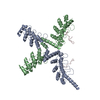
| ||||||||
|---|---|---|---|---|---|---|---|---|---|
| 1 |
| ||||||||
| Unit cell |
|
-Components
| #1: Protein | / TANK1 / TRF1-interacting ankyrin-related ADP-ribose polymerase 1 / Tankyrase I Mass: 38235.715 Da / Num. of mol.: 2 / Fragment: mTNKS1 ARC23 (UNP Residues 308-655) Source method: isolated from a genetically manipulated source Source: (gene. exp.) Mus musculus (house mouse) / Gene: TANKRYASE1, Tnks, Tnks1 / Plasmid: pGEX4T1 / Production host:  Escherichia coli (E. coli) / Strain (production host): BL21 / References: UniProt: Q6PFX9, NAD+ ADP-ribosyltransferase Escherichia coli (E. coli) / Strain (production host): BL21 / References: UniProt: Q6PFX9, NAD+ ADP-ribosyltransferase#2: Protein | | AXIN1 / Axis inhibition protein 1 / Protein FusedMass: 8784.486 Da / Num. of mol.: 1 / Fragment: mAxin1 N domain (UNP Residues 1-80) Source method: isolated from a genetically manipulated source Source: (gene. exp.) Mus musculus (house mouse) / Gene: Axin, Axin1, Fu / Plasmid: pAL / Production host: Escherichia coli (E. coli) / Strain (production host): BL21 / References: UniProt: O35625#3: Water | ChemComp-HOH / | Water Mass: 18.015 Da / Num. of mol.: 227 / Source method: isolated from a natural source / Formula: H2O Mass: 18.015 Da / Num. of mol.: 227 / Source method: isolated from a natural source / Formula: H2O |
|---|
-Experimental details
-Experiment
| Experiment | Method: X-RAY DIFFRACTION / Number of used crystals: 1 |
|---|
- Sample preparation
| Crystal | Density Matthews: 2.65 Å3/Da / Density % sol: 53.63 % |
|---|---|
| Crystal grow | Temperature: 295 K / Method: evaporation / pH: 5.6 Details: 0.06 M sodium citrate tribasic dihydrate pH 5.6, 1.2M ammonium acetate, 18% MPD, and 20mM DTT, EVAPORATION, temperature 295K |
-Data collection
| Diffraction | Mean temperature: 100 K |
|---|---|
| Diffraction source | Source: SYNCHROTRON / Site: ALS  / Beamline: 8.2.2 / Wavelength: 0.979 Å / Beamline: 8.2.2 / Wavelength: 0.979 Å |
| Detector | Type: ADSC QUANTUM 4 / Detector: CCD / Date: Nov 18, 2010 |
| Radiation | Protocol: SAD / Monochromatic (M) / Laue (L): M / Scattering type: x-ray |
| Radiation wavelength | Wavelength: 0.979 Å / Relative weight: 1 |
| Reflection | Resolution: 2→30 Å / Num. all: 70985 / Num. obs: 70726 / % possible obs: 99.85 % / Redundancy: 6.9 % / Rsym value: 0.059 / Net I/σ(I): 30 |
| Reflection shell | Resolution: 2→2.05 Å / Mean I/σ(I) obs: 2.7 / Rsym value: 0.66 |
- Processing
| Software |
| ||||||||||||||||||||||||||||||||||||||||||||||||||||||||||||||||||||||||||||||||||||||||||||||||||||||||||||||||||||||||||||||||||||||||||||||||||||||||||||||||||||||||||
|---|---|---|---|---|---|---|---|---|---|---|---|---|---|---|---|---|---|---|---|---|---|---|---|---|---|---|---|---|---|---|---|---|---|---|---|---|---|---|---|---|---|---|---|---|---|---|---|---|---|---|---|---|---|---|---|---|---|---|---|---|---|---|---|---|---|---|---|---|---|---|---|---|---|---|---|---|---|---|---|---|---|---|---|---|---|---|---|---|---|---|---|---|---|---|---|---|---|---|---|---|---|---|---|---|---|---|---|---|---|---|---|---|---|---|---|---|---|---|---|---|---|---|---|---|---|---|---|---|---|---|---|---|---|---|---|---|---|---|---|---|---|---|---|---|---|---|---|---|---|---|---|---|---|---|---|---|---|---|---|---|---|---|---|---|---|---|---|---|---|---|---|
| Refinement | Method to determine structure: MOLECULAR REPLACEMENT Starting model: PDB ENTRY 1YMP Resolution: 2→30 Å / Cor.coef. Fo:Fc: 0.963 / Cor.coef. Fo:Fc free: 0.948 / Occupancy max: 1 / Occupancy min: 1 / SU B: 7.628 / SU ML: 0.117 / Cross valid method: THROUGHOUT / ESU R: 0.142 / ESU R Free: 0.141 / Stereochemistry target values: MAXIMUM LIKELIHOOD / Details: HYDROGENS HAVE BEEN USED IF PRESENT IN THE INPUT
| ||||||||||||||||||||||||||||||||||||||||||||||||||||||||||||||||||||||||||||||||||||||||||||||||||||||||||||||||||||||||||||||||||||||||||||||||||||||||||||||||||||||||||
| Solvent computation | Ion probe radii: 0.8 Å / Shrinkage radii: 0.8 Å / VDW probe radii: 1.2 Å / Solvent model: MASK | ||||||||||||||||||||||||||||||||||||||||||||||||||||||||||||||||||||||||||||||||||||||||||||||||||||||||||||||||||||||||||||||||||||||||||||||||||||||||||||||||||||||||||
| Displacement parameters | Biso mean: 63.174 Å2
| ||||||||||||||||||||||||||||||||||||||||||||||||||||||||||||||||||||||||||||||||||||||||||||||||||||||||||||||||||||||||||||||||||||||||||||||||||||||||||||||||||||||||||
| Refinement step | Cycle: LAST / Resolution: 2→30 Å
| ||||||||||||||||||||||||||||||||||||||||||||||||||||||||||||||||||||||||||||||||||||||||||||||||||||||||||||||||||||||||||||||||||||||||||||||||||||||||||||||||||||||||||
| Refine LS restraints |
| ||||||||||||||||||||||||||||||||||||||||||||||||||||||||||||||||||||||||||||||||||||||||||||||||||||||||||||||||||||||||||||||||||||||||||||||||||||||||||||||||||||||||||
| LS refinement shell | Resolution: 2→2.052 Å / Total num. of bins used: 20
| ||||||||||||||||||||||||||||||||||||||||||||||||||||||||||||||||||||||||||||||||||||||||||||||||||||||||||||||||||||||||||||||||||||||||||||||||||||||||||||||||||||||||||
| Refinement TLS params. | Method: refined / Refine-ID: X-RAY DIFFRACTION
| ||||||||||||||||||||||||||||||||||||||||||||||||||||||||||||||||||||||||||||||||||||||||||||||||||||||||||||||||||||||||||||||||||||||||||||||||||||||||||||||||||||||||||
| Refinement TLS group |
|
