 Movie
Movie Controller


+ Open data

- Basic information
| Entry | Database: PDB / ID: 5dsy | ||||||
|---|---|---|---|---|---|---|---|
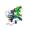
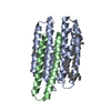
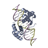
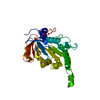
| Title | Crystal structure of constitutively active PARP-2 | ||||||
 Components Components | Poly [ADP-ribose] polymerase 2 | ||||||
 Keywords Keywords |  TRANSFERASE / ADP-ribosyl transferase / PARP / PARP-2 TRANSFERASE / ADP-ribosyl transferase / PARP / PARP-2 | ||||||
| Function / homology |  Function and homology information Function and homology informationhippocampal neuron apoptotic process / response to oxygen-glucose deprivation / poly-ADP-D-ribose binding / positive regulation of cell growth involved in cardiac muscle cell development / NAD+-protein-serine ADP-ribosyltransferase activity / NAD DNA ADP-ribosyltransferase activity / NAD+- protein-aspartate ADP-ribosyltransferase activity / NAD+-protein-glutamate ADP-ribosyltransferase activity / DNA ADP-ribosylation / HDR through MMEJ (alt-NHEJ) ...hippocampal neuron apoptotic process / response to oxygen-glucose deprivation / poly-ADP-D-ribose binding / positive regulation of cell growth involved in cardiac muscle cell development / NAD+-protein-serine ADP-ribosyltransferase activity / NAD DNA ADP-ribosyltransferase activity / NAD+- protein-aspartate ADP-ribosyltransferase activity / NAD+-protein-glutamate ADP-ribosyltransferase activity / DNA ADP-ribosylation / HDR through MMEJ (alt-NHEJ) / poly-ADP-D-ribose modification-dependent protein binding / DNA repair-dependent chromatin remodeling / NAD+ ADP-ribosyltransferase / protein auto-ADP-ribosylation / protein poly-ADP-ribosylation / site of DNA damage / NAD+-protein ADP-ribosyltransferase activity / decidualization / Transferases; Glycosyltransferases; Pentosyltransferases / NAD+ ADP-ribosyltransferase activity / POLB-Dependent Long Patch Base Excision Repair / nucleosome binding / extrinsic apoptotic signaling pathway / nucleotidyltransferase activity / DNA Damage Recognition in GG-NER / base-excision repair / Dual Incision in GG-NER / Formation of Incision Complex in GG-NER / double-strand break repair / damaged DNA binding / DNA repair / DNA damage response / chromatin binding / nucleolus / nucleoplasm / nucleusSimilarity search - Function | ||||||
| Biological species |  Homo sapiens (human) Homo sapiens (human) | ||||||
| Method | X-RAY DIFFRACTION / SYNCHROTRON / MOLECULAR REPLACEMENT / Resolution: 2.7 Å | ||||||
 Authors Authors | Riccio, A.A. / Pascal, J.M. | ||||||
| Funding support |  United States, 1items United States, 1items
| ||||||
 Citation Citation | Journal: Mol.Cell / Year: 2015 Title: PARP-1 Activation Requires Local Unfolding of an Autoinhibitory Domain. Authors: Dawicki-McKenna, J.M. / Langelier, M.F. / DeNizio, J.E. / Riccio, A.A. / Cao, C.D. / Karch, K.R. / McCauley, M. / Steffen, J.D. / Black, B.E. / Pascal, J.M. | ||||||
| History |
|
- Structure visualization
| Structure viewer | Molecule: MolmilJmol/JSmol |
|---|
- Downloads & links
-Download
| PDBx/mmCIF format | 5dsy.cif.gz | 210.6 KB | Display | PDBx/mmCIF format |
|---|---|---|---|---|
| PDB format | pdb5dsy.ent.gz | 168.3 KB | Display | PDB format |
| PDBx/mmJSON format | 5dsy.json.gz | Tree view | PDBx/mmJSON format | |
| Others |  Other downloads Other downloads |
-Validation report
| Arichive directory | https://data.pdbj.org/pub/pdb/validation_reports/ds/5dsyftp://data.pdbj.org/pub/pdb/validation_reports/ds/5dsy | HTTPS FTP |
|---|
-Related structure data
| Related structure data | 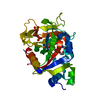 5ds3C  3kjdS S: Starting model for refinement C: citing same article ( |
|---|---|
| Similar structure data |


-Links
 PDBj
PDBj

- Assembly
| Deposited unit | 
| ||||||||
|---|---|---|---|---|---|---|---|---|---|
| 1 | 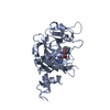
| ||||||||
| 2 | 
| ||||||||
| 3 | 
| ||||||||
| 4 | 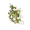
| ||||||||
| Unit cell |
|
-Components
| #1: Protein | Mass: 31675.916 Da / Num. of mol.: 4 / Fragment: unp residues 348-583 Source method: isolated from a genetically manipulated source Source: (gene. exp.) Homo sapiens (human) / Gene: PARP2, ADPRT2, ADPRTL2 / Production host:  Escherichia coli (E. coli) / References: UniProt: Q9UGN5, NAD+ ADP-ribosyltransferase Escherichia coli (E. coli) / References: UniProt: Q9UGN5, NAD+ ADP-ribosyltransferase#2: Chemical | ChemComp-UHB /   Mass: 537.528 Da / Num. of mol.: 4 / Source method: obtained synthetically / Formula: C24H27N9O6 Mass: 537.528 Da / Num. of mol.: 4 / Source method: obtained synthetically / Formula: C24H27N9O6#3: Water | ChemComp-HOH / | Water Mass: 18.015 Da / Num. of mol.: 51 / Source method: isolated from a natural source / Formula: H2O Mass: 18.015 Da / Num. of mol.: 51 / Source method: isolated from a natural source / Formula: H2O |
|---|
-Experimental details
-Experiment
| Experiment | Method: X-RAY DIFFRACTION |
|---|
- Sample preparation
| Crystal | Density Matthews: 2.63 Å3/Da / Density % sol: 53.28 % |
|---|---|
| Crystal grow | Temperature: 298 K / Method: vapor diffusion, sitting drop / pH: 8 / Details: 2.55-2.65 M NaCl and 0.1 M Tris / PH range: 7.5-8.5 |
-Data collection
| Diffraction | Mean temperature: 100 K |
|---|---|
| Diffraction source | Source: SYNCHROTRON / Site: ALS / Beamline: 12.3.1 / Wavelength: 1.12 Å |
| Detector | Type: ADSC QUANTUM 315r / Detector: CCD / Date: Aug 29, 2014 |
| Radiation | Protocol: SINGLE WAVELENGTH / Monochromatic (M) / Laue (L): M / Scattering type: x-ray |
| Radiation wavelength | Wavelength: 1.12 Å / Relative weight: 1 |
| Reflection | Resolution: 2.7→50 Å / Num. obs: 70646 / % possible obs: 98.4 % / Redundancy: 12 % / Net I/σ(I): 11 |
| Reflection shell | Resolution: 2.68→2.8 Å / Redundancy: 11.5 % / Mean I/σ(I) obs: 1.3 / % possible all: 87.2 |
- Processing
| Software |
| ||||||||||||||||||||||||||||||||||||||||||||||||||||||||||||||||||||||||||||||||||||||||||||||||||||||||||||||||||||||||||||||||||||||||||||||||||||||||||||||||||||||||||||||||||||||
|---|---|---|---|---|---|---|---|---|---|---|---|---|---|---|---|---|---|---|---|---|---|---|---|---|---|---|---|---|---|---|---|---|---|---|---|---|---|---|---|---|---|---|---|---|---|---|---|---|---|---|---|---|---|---|---|---|---|---|---|---|---|---|---|---|---|---|---|---|---|---|---|---|---|---|---|---|---|---|---|---|---|---|---|---|---|---|---|---|---|---|---|---|---|---|---|---|---|---|---|---|---|---|---|---|---|---|---|---|---|---|---|---|---|---|---|---|---|---|---|---|---|---|---|---|---|---|---|---|---|---|---|---|---|---|---|---|---|---|---|---|---|---|---|---|---|---|---|---|---|---|---|---|---|---|---|---|---|---|---|---|---|---|---|---|---|---|---|---|---|---|---|---|---|---|---|---|---|---|---|---|---|---|---|
| Refinement | Method to determine structure: MOLECULAR REPLACEMENT Starting model: 3KJD Resolution: 2.7→46.54 Å / SU ML: 0.38 / Cross valid method: THROUGHOUT / σ(F): 1.32 / Phase error: 26.13 / Stereochemistry target values: ML
| ||||||||||||||||||||||||||||||||||||||||||||||||||||||||||||||||||||||||||||||||||||||||||||||||||||||||||||||||||||||||||||||||||||||||||||||||||||||||||||||||||||||||||||||||||||||
| Solvent computation | Shrinkage radii: 0.9 Å / VDW probe radii: 1.11 Å / Solvent model: FLAT BULK SOLVENT MODEL | ||||||||||||||||||||||||||||||||||||||||||||||||||||||||||||||||||||||||||||||||||||||||||||||||||||||||||||||||||||||||||||||||||||||||||||||||||||||||||||||||||||||||||||||||||||||
| Refinement step | Cycle: LAST / Resolution: 2.7→46.54 Å
| ||||||||||||||||||||||||||||||||||||||||||||||||||||||||||||||||||||||||||||||||||||||||||||||||||||||||||||||||||||||||||||||||||||||||||||||||||||||||||||||||||||||||||||||||||||||
| Refine LS restraints |
| ||||||||||||||||||||||||||||||||||||||||||||||||||||||||||||||||||||||||||||||||||||||||||||||||||||||||||||||||||||||||||||||||||||||||||||||||||||||||||||||||||||||||||||||||||||||
| LS refinement shell |
|
