 Movie
Movie Controller

[English] 日本語
 Yorodumi
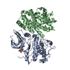
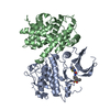
- PDB-5jje: Structure of the SRII/HtrII Complex in I212121 space group ("U" shape) -
+ Open data

- Basic information
| Entry | Database: PDB / ID: 5jje | |||||||||
|---|---|---|---|---|---|---|---|---|---|---|
| Title | Structure of the SRII/HtrII Complex in I212121 space group ("U" shape) | |||||||||
 Components Components |
| |||||||||
 Keywords Keywords |  SIGNALING PROTEIN / sensory rhodopsin II / transducer / membrane protein complex SIGNALING PROTEIN / sensory rhodopsin II / transducer / membrane protein complex | |||||||||
| Function / homology |  Function and homology informationphotoreceptor activity / phototransduction / transmembrane signaling receptor activity / chemotaxis / lysozyme activity / monoatomic ion channel activity / signal transduction / identical protein binding / plasma membrane Function and homology informationphotoreceptor activity / phototransduction / transmembrane signaling receptor activity / chemotaxis / lysozyme activity / monoatomic ion channel activity / signal transduction / identical protein binding / plasma membraneSimilarity search - Function | |||||||||
| Biological species |  Natronomonas pharaonis (archaea) Natronomonas pharaonis (archaea) | |||||||||
| Method | X-RAY DIFFRACTION / SYNCHROTRON / MOLECULAR REPLACEMENT / Resolution: 1.9 Å | |||||||||
 Authors Authors | Ishchenko, A. / Round, E. / Borshchevskiy, V. / Grudinin, S. / Gushchin, I. / Klare, J. / Remeeva, A. / Polovinkin, V. / Utrobin, P. / Balandin, T. ...Ishchenko, A. / Round, E. / Borshchevskiy, V. / Grudinin, S. / Gushchin, I. / Klare, J. / Remeeva, A. / Polovinkin, V. / Utrobin, P. / Balandin, T. / Engelhard, M. / Bueldt, G. / Gordeliy, V. | |||||||||
| Funding support |  Russian Federation, Russian Federation,  France, 2items France, 2items
| |||||||||
 Citation Citation | Journal: Sci Rep / Year: 2017 Title: New Insights on Signal Propagation by Sensory Rhodopsin II/Transducer Complex. Authors: Ishchenko, A. / Round, E. / Borshchevskiy, V. / Grudinin, S. / Gushchin, I. / Klare, J.P. / Remeeva, A. / Polovinkin, V. / Utrobin, P. / Balandin, T. / Engelhard, M. / Buldt, G. / Gordeliy, V. | |||||||||
| History |
|
- Structure visualization
| Structure viewer | Molecule: MolmilJmol/JSmol |
|---|
- Downloads & links
-Download
| PDBx/mmCIF format | 5jje.cif.gz | 125.4 KB | Display | PDBx/mmCIF format |
|---|---|---|---|---|
| PDB format | pdb5jje.ent.gz | 93.8 KB | Display | PDB format |
| PDBx/mmJSON format | 5jje.json.gz | Tree view | PDBx/mmJSON format | |
| Others |  Other downloads Other downloads |
-Validation report
| Arichive directory | https://data.pdbj.org/pub/pdb/validation_reports/jj/5jjeftp://data.pdbj.org/pub/pdb/validation_reports/jj/5jje | HTTPS FTP |
|---|
-Related structure data
| Related structure data | 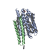 5jjfC  5jjjC 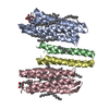 5jjnC 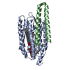 1h2sS S: Starting model for refinement C: citing same article ( |
|---|---|
| Similar structure data |


-Links
 PDBj
PDBj


- Assembly
| Deposited unit | 
| ||||||||
|---|---|---|---|---|---|---|---|---|---|
| 1 | 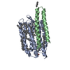
| ||||||||
| Unit cell |
|
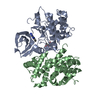
-Components
-Protein , 2 types, 2 molecules AB
| #1: Protein | Mass: 26534.941 Da / Num. of mol.: 1 Source method: isolated from a genetically manipulated source Source: (gene. exp.) Natronomonas pharaonis (archaea) / Gene: sop2, sopII / Production host:  Escherichia coli (E. coli) / References: UniProt: P42196 Escherichia coli (E. coli) / References: UniProt: P42196 |
|---|---|
| #2: Protein | Mass: 17224.270 Da / Num. of mol.: 1 Source method: isolated from a genetically manipulated source Source: (gene. exp.) Natronomonas pharaonis (archaea) / Gene: htr2, htrII / Production host: Escherichia coli (E. coli) / References: UniProt: P42259 |
-Sugars , 1 types, 1 molecules 
| #4: Sugar | ChemComp-BOG / Octyl glucoside Type: D-saccharide / Mass: 292.369 Da / Num. of mol.: 1 Type: D-saccharide / Mass: 292.369 Da / Num. of mol.: 1Source method: isolated from a genetically manipulated source Formula: C14H28O6 / Comment: detergent*YM |
|---|
-Non-polymers , 3 types, 69 molecules 


| #3: Chemical | ChemComp-RET / Retinal Mass: 284.436 Da / Num. of mol.: 1 / Source method: obtained synthetically / Formula: C20H28O Mass: 284.436 Da / Num. of mol.: 1 / Source method: obtained synthetically / Formula: C20H28O | ||
|---|---|---|---|
| #5: Chemical | ChemComp-LFA / Icosane Mass: 282.547 Da / Num. of mol.: 12 / Source method: obtained synthetically / Formula: C20H42 Mass: 282.547 Da / Num. of mol.: 12 / Source method: obtained synthetically / Formula: C20H42#6: Water | ChemComp-HOH / | WaterMass: 18.015 Da / Num. of mol.: 56 / Source method: isolated from a natural source / Formula: H2O |
-Experimental details
-Experiment
| Experiment | Method: X-RAY DIFFRACTION / Number of used crystals: 1 |
|---|
- Sample preparation
| Crystal | Density Matthews: 2.03 Å3/Da / Density % sol: 39.43 % |
|---|---|
| Crystal grow | Temperature: 293 K / Method: lipidic cubic phase / Details: 1 M Na/K phosphate, pH 5.6 |
-Data collection
| Diffraction | Mean temperature: 100 K |
|---|---|
| Diffraction source | Source: SYNCHROTRON / Site: ESRF / Beamline: ID14-1 / Wavelength: 0.987 Å |
| Detector | Type: ADSC QUANTUM 210 / Detector: CCD / Date: Oct 20, 2009 |
| Radiation | Protocol: SINGLE WAVELENGTH / Monochromatic (M) / Laue (L): M / Scattering type: x-ray |
| Radiation wavelength | Wavelength: 0.987 Å / Relative weight: 1 |
| Reflection | Resolution: 1.9→28.1348 Å / Num. obs: 28430 / % possible obs: 99.4 % / Redundancy: 3.8 % / Rmerge(I) obs: 0.07 / Net I/σ(I): 13.4 |
| Reflection shell | Resolution: 1.9→2 Å / Rmerge(I) obs: 0.393 |
- Processing
| Software |
| |||||||||||||||||||||||||||||||||||||||||||||||||||||||||||||||||||||||||||||
|---|---|---|---|---|---|---|---|---|---|---|---|---|---|---|---|---|---|---|---|---|---|---|---|---|---|---|---|---|---|---|---|---|---|---|---|---|---|---|---|---|---|---|---|---|---|---|---|---|---|---|---|---|---|---|---|---|---|---|---|---|---|---|---|---|---|---|---|---|---|---|---|---|---|---|---|---|---|---|
| Refinement | Method to determine structure: MOLECULAR REPLACEMENT Starting model: 1h2s Resolution: 1.9→28.1348 Å / Cross valid method: FREE R-VALUE
| |||||||||||||||||||||||||||||||||||||||||||||||||||||||||||||||||||||||||||||
| Refinement step | Cycle: LAST / Resolution: 1.9→28.1348 Å
| |||||||||||||||||||||||||||||||||||||||||||||||||||||||||||||||||||||||||||||
| Refine LS restraints |
| |||||||||||||||||||||||||||||||||||||||||||||||||||||||||||||||||||||||||||||
| LS refinement shell |
|
