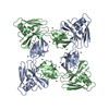
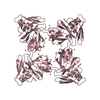
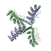
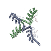
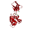
Entry Database : PDB / ID : 5xljTitle Crystal structure of the flagellar cap protein flid D2-D3 domains from serratia marcescens in Space group P432 Flagellar hook-associated protein 2 Keywords / Function / homology Function Domain/homology Component
/ / / / / / Biological species Serratia marcescens (bacteria)Method / / / Resolution : 1.9 Å Authors Cho, S.Y. / Song, W.S. / Hong, H.J. / Yoon, S.I. Journal : Biochem. Biophys. Res. Commun. / Year : 2017Title : Tetrameric structure of the flagellar cap protein FliD from Serratia marcescens.Authors : Cho, S.Y. / Song, W.S. / Hong, H.J. / Lee, G.S. / Kang, S.G. / Ko, H.J. / Kim, P.H. / Yoon, S.I. History Deposition May 10, 2017 Deposition site / Processing site Revision 1.0 Jun 14, 2017 Provider / Type Revision 1.1 Dec 6, 2017 Group / Category Item / _citation.page_first / _citation.page_lastRevision 1.2 Nov 22, 2023 Group Data collection / Database references ... Data collection / Database references / Derived calculations / Refinement description Category chem_comp_atom / chem_comp_bond ... chem_comp_atom / chem_comp_bond / database_2 / pdbx_initial_refinement_model / pdbx_struct_conn_angle / struct_conn Item _database_2.pdbx_DOI / _database_2.pdbx_database_accession ... _database_2.pdbx_DOI / _database_2.pdbx_database_accession / _pdbx_struct_conn_angle.ptnr1_auth_comp_id / _pdbx_struct_conn_angle.ptnr1_auth_seq_id / _pdbx_struct_conn_angle.ptnr1_label_asym_id / _pdbx_struct_conn_angle.ptnr1_label_comp_id / _pdbx_struct_conn_angle.ptnr1_label_seq_id / _pdbx_struct_conn_angle.ptnr1_symmetry / _pdbx_struct_conn_angle.ptnr2_symmetry / _pdbx_struct_conn_angle.ptnr3_auth_comp_id / _pdbx_struct_conn_angle.ptnr3_auth_seq_id / _pdbx_struct_conn_angle.ptnr3_label_asym_id / _pdbx_struct_conn_angle.ptnr3_label_comp_id / _pdbx_struct_conn_angle.ptnr3_label_seq_id / _pdbx_struct_conn_angle.ptnr3_symmetry / _pdbx_struct_conn_angle.value / _struct_conn.pdbx_dist_value / _struct_conn.ptnr1_auth_comp_id / _struct_conn.ptnr1_auth_seq_id / _struct_conn.ptnr1_label_asym_id / _struct_conn.ptnr1_label_atom_id / _struct_conn.ptnr1_label_comp_id / _struct_conn.ptnr1_label_seq_id / _struct_conn.ptnr2_auth_comp_id / _struct_conn.ptnr2_auth_seq_id / _struct_conn.ptnr2_label_asym_id / _struct_conn.ptnr2_label_atom_id / _struct_conn.ptnr2_label_comp_id / _struct_conn.ptnr2_symmetry
Show all Show less
 Movie
Movie Controller

 Yorodumi
 Open data
 Basic information
 Components
 Keywords
 STRUCTURAL PROTEIN / Bacterial flagellar cap protein
 Function and homology information
 Authors
 Citation
 Structure visualization
 Downloads & links
 Other downloads


 PDBj
PDBj Assembly
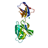

 Mass: 35.453 Da / Num. of mol.: 5 / Source method: obtained synthetically / Formula: Cl
 Mass: 22.990 Da / Num. of mol.: 1 / Source method: obtained synthetically / Formula: Na
 Mass: 18.015 Da / Num. of mol.: 150 / Source method: isolated from a natural source / Formula: H2O
 Sample preparation
 / Beamline: 7A (6B, 6C1) / Wavelength: 1.00004 Å
 Processing