 Movie
Movie Controller


+ Open data

- Basic information
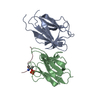
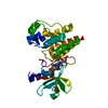
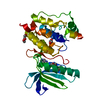
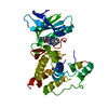
| Entry | Database: PDB / ID: 3rk6 | ||||||
|---|---|---|---|---|---|---|---|
| Title | Crystal structure of the middle domain of human Paip1 | ||||||
 Components Components | Polyadenylate-binding protein-interacting protein 1 | ||||||
 Keywords Keywords | TRANSLATION REGULATOR / HEAT Fold /  PABP / eIF4A / eIF3 PABP / eIF4A / eIF3 | ||||||
| Function / homology |  Function and homology information Function and homology informationmCRD-mediated mRNA stability complex / translation activator activity / negative regulation of nuclear-transcribed mRNA catabolic process, deadenylation-dependent decay / CRD-mediated mRNA stabilization / Z-decay: degradation of maternal mRNAs by zygotically expressed factors / Deadenylation of mRNA / positive regulation of cytoplasmic translation / positive regulation by host of viral process / mRNA stabilization / M-decay: degradation of maternal mRNAs by maternally stored factors ...mCRD-mediated mRNA stability complex / translation activator activity / negative regulation of nuclear-transcribed mRNA catabolic process, deadenylation-dependent decay / CRD-mediated mRNA stabilization / Z-decay: degradation of maternal mRNAs by zygotically expressed factors / Deadenylation of mRNA / positive regulation of cytoplasmic translation / positive regulation by host of viral process / mRNA stabilization / M-decay: degradation of maternal mRNAs by maternally stored factors / regulation of translational initiation / translational initiation / viral translation / RNA binding / cytosol / cytoplasmSimilarity search - Function | ||||||
| Biological species |  Homo sapiens (human) Homo sapiens (human) | ||||||
| Method | X-RAY DIFFRACTION / SYNCHROTRON / SAD / Resolution: 2 Å | ||||||
 Authors Authors | Lei, J. / Mesters, J.R. / Hilgenfeld, R. | ||||||
 Citation Citation | Journal: Biochem.Biophys.Res.Commun. / Year: 2011 Title: Crystal structure of the middle domain of human poly(A)-binding protein-interacting protein 1. Authors: Lei, J. / Mesters, J.R. / Brunn, A. / Hilgenfeld, R. | ||||||
| History |
|
- Structure visualization
| Structure viewer | Molecule: MolmilJmol/JSmol |
|---|
- Downloads & links
-Download
| PDBx/mmCIF format | 3rk6.cif.gz | 107.5 KB | Display | PDBx/mmCIF format |
|---|---|---|---|---|
| PDB format | pdb3rk6.ent.gz | 83.4 KB | Display | PDB format |
| PDBx/mmJSON format | 3rk6.json.gz | Tree view | PDBx/mmJSON format | |
| Others |  Other downloads Other downloads |
-Validation report
| Arichive directory | https://data.pdbj.org/pub/pdb/validation_reports/rk/3rk6ftp://data.pdbj.org/pub/pdb/validation_reports/rk/3rk6 | HTTPS FTP |
|---|
-Related structure data
| Similar structure data |
|---|


-Links
 PDBj
PDBj


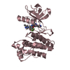
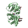
- Assembly
| Deposited unit | 
| ||||||||
|---|---|---|---|---|---|---|---|---|---|
| 1 | 
| ||||||||
| 2 | 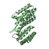
| ||||||||
| Unit cell |
|
-Components
| #1: Protein | Mass: 26480.188 Da / Num. of mol.: 2 / Fragment: middle domain (MIF4G, UNP residues 157-373) Source method: isolated from a genetically manipulated source Source: (gene. exp.) Homo sapiens (human) / Gene: PAIP1 / Production host:  Escherichia coli (E. coli) / References: UniProt: Q9H074 Escherichia coli (E. coli) / References: UniProt: Q9H074#2: Water | ChemComp-HOH / | Water Mass: 18.015 Da / Num. of mol.: 370 / Source method: isolated from a natural source / Formula: H2O Mass: 18.015 Da / Num. of mol.: 370 / Source method: isolated from a natural source / Formula: H2O |
|---|
-Experimental details
-Experiment
| Experiment | Method: X-RAY DIFFRACTION / Number of used crystals: 1 |
|---|
- Sample preparation
| Crystal | Density Matthews: 2.61 Å3/Da / Density % sol: 52.87 % |
|---|---|
| Crystal grow | Temperature: 293 K / Method: vapor diffusion, sitting drop / pH: 6.5 Details: 18% PEG20000, 0.1 M MES, pH 6.5, VAPOR DIFFUSION, SITTING DROP, temperature 293K |
-Data collection
| Diffraction | Mean temperature: 90 K |
|---|---|
| Diffraction source | Source: SYNCHROTRON / Site: EMBL/DESY, HAMBURG  / Beamline: X11 / Wavelength: 0.817 / Beamline: X11 / Wavelength: 0.817 |
| Detector | Type: MAR scanner 300 mm plate / Detector: IMAGE PLATE / Date: Nov 19, 2010 |
| Radiation | Monochromator: Ge(111) triangular bent compressing 7 degree Fankuchen cut Protocol: SINGLE WAVELENGTH / Monochromatic (M) / Laue (L): M / Scattering type: x-ray |
| Radiation wavelength | Wavelength: 0.817 Å / Relative weight: 1 |
| Reflection | Resolution: 2→19.95 Å / Num. all: 36850 / Num. obs: 36673 / % possible obs: 99.6 % / Observed criterion σ(F): 2 / Observed criterion σ(I): 2 / Redundancy: 3.8 % / Rmerge(I) obs: 0.049 / Rsym value: 0.029 / Net I/σ(I): 14.9 |
| Reflection shell | Resolution: 2→2.11 Å / Redundancy: 3.8 % / Rmerge(I) obs: 0.023 / Mean I/σ(I) obs: 5.1 / Num. unique all: 5348 / Rsym value: 0.136 / % possible all: 100 |
-Phasing
| Phasing | Method: SAD |
|---|
- Processing
| Software |
| |||||||||||||||||||||||||||||||||||||||||||||||||||||||||||||||||
|---|---|---|---|---|---|---|---|---|---|---|---|---|---|---|---|---|---|---|---|---|---|---|---|---|---|---|---|---|---|---|---|---|---|---|---|---|---|---|---|---|---|---|---|---|---|---|---|---|---|---|---|---|---|---|---|---|---|---|---|---|---|---|---|---|---|---|
| Refinement | Method to determine structure: SAD / Resolution: 2→19.95 Å / Cor.coef. Fo:Fc: 0.958 / Cor.coef. Fo:Fc free: 0.923 / WRfactor Rfree: 0.2413 / WRfactor Rwork: 0.1887 / Occupancy max: 1 / Occupancy min: 0.5 / FOM work R set: 0.8017 / SU B: 3.791 / SU ML: 0.108 / SU R Cruickshank DPI: 0.1761 / SU Rfree: 0.1673 / Cross valid method: THROUGHOUT / σ(F): 0 / ESU R: 0.176 / ESU R Free: 0.167 / Stereochemistry target values: MAXIMUM LIKELIHOOD Details: HYDROGENS HAVE BEEN ADDED IN THE RIDING POSITIONS U VALUES : REFINED INDIVIDUALLY
| |||||||||||||||||||||||||||||||||||||||||||||||||||||||||||||||||
| Solvent computation | Ion probe radii: 0.8 Å / Shrinkage radii: 0.8 Å / VDW probe radii: 1.4 Å / Solvent model: MASK | |||||||||||||||||||||||||||||||||||||||||||||||||||||||||||||||||
| Displacement parameters | Biso max: 91.64 Å2 / Biso mean: 27.5042 Å2 / Biso min: 11.45 Å2
| |||||||||||||||||||||||||||||||||||||||||||||||||||||||||||||||||
| Refinement step | Cycle: LAST / Resolution: 2→19.95 Å
| |||||||||||||||||||||||||||||||||||||||||||||||||||||||||||||||||
| Refine LS restraints |
| |||||||||||||||||||||||||||||||||||||||||||||||||||||||||||||||||
| LS refinement shell | Resolution: 2→2.051 Å / Total num. of bins used: 20
|
