 Movie
Movie Controller

[English] 日本語
 Yorodumi
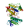
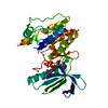
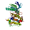
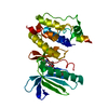
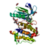
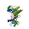
- PDB-3wyx: CRYSTAL STRUCTURE OF HUMAN MPS1 CATALYTIC DOMAIN IN COMPLEX WITH ... -
+ Open data

- Basic information
| Entry | Database: PDB / ID: 3wyx | ||||||
|---|---|---|---|---|---|---|---|
| Title | CRYSTAL STRUCTURE OF HUMAN MPS1 CATALYTIC DOMAIN IN COMPLEX WITH 6-((3-(cyanomethoxy)-4-(1-methyl-1H-pyrazol-4-yl)phenyl)amino)-2-(cyclohexylamino)nicotinonitrile | ||||||
 Components Components | Dual specificity protein kinase TTK | ||||||
 Keywords Keywords |  TRANSFERASE / KINASE / ATP Binding TRANSFERASE / KINASE / ATP Binding | ||||||
| Function / homology |  Function and homology information Function and homology informationprotein localization to meiotic spindle midzone / meiotic spindle assembly checkpoint signaling / kinetochore binding / female meiosis chromosome segregation / protein localization to kinetochore / dual-specificity kinase / spindle organization / mitotic spindle assembly checkpoint signaling / protein serine/threonine/tyrosine kinase activity / mitotic spindle organization ...protein localization to meiotic spindle midzone / meiotic spindle assembly checkpoint signaling / kinetochore binding / female meiosis chromosome segregation / protein localization to kinetochore / dual-specificity kinase / spindle organization / mitotic spindle assembly checkpoint signaling / protein serine/threonine/tyrosine kinase activity / mitotic spindle organization / chromosome segregation / kinetochore / spindle / protein tyrosine kinase activity / phosphorylation / protein serine kinase activity / protein serine/threonine kinase activity / positive regulation of cell population proliferation / ATP binding / membrane / identical protein binding / nucleus / cytoplasmSimilarity search - Function | ||||||
| Biological species |  Homo sapiens (human) Homo sapiens (human) | ||||||
| Method | X-RAY DIFFRACTION / MOLECULAR REPLACEMENT / molecular replacement / Resolution: 2.9 Å | ||||||
 Authors Authors | Kusakabe, K. / Ide, N. / Daigo, Y. / Itoh, T. / Yamamoto, T. / Kojima, E. / Mitsuoka, Y. / Tadano, G. / Tagashira, S. / Higashino, K. ...Kusakabe, K. / Ide, N. / Daigo, Y. / Itoh, T. / Yamamoto, T. / Kojima, E. / Mitsuoka, Y. / Tadano, G. / Tagashira, S. / Higashino, K. / Okano, Y. / Sato, Y. / Inoue, M. / Iguchi, M. / Kanazawa, T. / Ishioka, Y. / Dohi, K. / Kido, Y. / Sakamoto, S. / Ando, S. / Maeda, M. / Higaki, M. / Yoshizawa, H. / Mura, H. / Nakamura, Y. | ||||||
 Citation Citation | Journal: Bioorg.Med.Chem. / Year: 2015 Title: A unique hinge binder of extremely selective aminopyridine-based Mps1 (TTK) kinase inhibitors with cellular activity. Authors: Kusakabe, K. / Ide, N. / Daigo, Y. / Itoh, T. / Yamamoto, T. / Kojima, E. / Mitsuoka, Y. / Tadano, G. / Tagashira, S. / Higashino, K. / Okano, Y. / Sato, Y. / Inoue, M. / Iguchi, M. / ...Authors: Kusakabe, K. / Ide, N. / Daigo, Y. / Itoh, T. / Yamamoto, T. / Kojima, E. / Mitsuoka, Y. / Tadano, G. / Tagashira, S. / Higashino, K. / Okano, Y. / Sato, Y. / Inoue, M. / Iguchi, M. / Kanazawa, T. / Ishioka, Y. / Dohi, K. / Kido, Y. / Sakamoto, S. / Ando, S. / Maeda, M. / Higaki, M. / Yoshizawa, H. / Murai, H. / Nakamura, Y. | ||||||
| History |
|
- Structure visualization
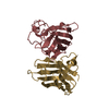
| Structure viewer | Molecule: MolmilJmol/JSmol |
|---|
- Downloads & links
-Download
| PDBx/mmCIF format | 3wyx.cif.gz | 67.3 KB | Display | PDBx/mmCIF format |
|---|---|---|---|---|
| PDB format | pdb3wyx.ent.gz | 47.3 KB | Display | PDB format |
| PDBx/mmJSON format | 3wyx.json.gz | Tree view | PDBx/mmJSON format | |
| Others |  Other downloads Other downloads |
-Validation report
| Arichive directory | https://data.pdbj.org/pub/pdb/validation_reports/wy/3wyxftp://data.pdbj.org/pub/pdb/validation_reports/wy/3wyx | HTTPS FTP |
|---|
-Related structure data


-Links
 PDBj
PDBj- Assembly
| Deposited unit | 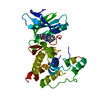
| ||||||||
|---|---|---|---|---|---|---|---|---|---|
| 1 |
| ||||||||
| 2 | 
| ||||||||
| Unit cell |
|
-Components
| #1: Protein | Mass: 36858.363 Da / Num. of mol.: 1 / Fragment: Mps1 (TTK) Kinase, UNP residues 516-820 Source method: isolated from a genetically manipulated source Source: (gene. exp.) Homo sapiens (human) / Gene: TTK, MPS1, MPS1L1 / Production host:  Escherichia coli (E. coli) / References: UniProt: P33981, dual-specificity kinase Escherichia coli (E. coli) / References: UniProt: P33981, dual-specificity kinase |
|---|---|
| #2: Chemical | ChemComp-IOD / Iodide  Mass: 126.904 Da / Num. of mol.: 1 / Source method: obtained synthetically / Formula: I Mass: 126.904 Da / Num. of mol.: 1 / Source method: obtained synthetically / Formula: I |
| #3: Chemical | ChemComp-O38 /   Mass: 427.502 Da / Num. of mol.: 1 / Source method: obtained synthetically / Formula: C24H25N7O Mass: 427.502 Da / Num. of mol.: 1 / Source method: obtained synthetically / Formula: C24H25N7O |
| #4: Water | ChemComp-HOH / Water Mass: 18.015 Da / Num. of mol.: 5 / Source method: isolated from a natural source / Formula: H2O Mass: 18.015 Da / Num. of mol.: 5 / Source method: isolated from a natural source / Formula: H2O |
-Experimental details
-Experiment
| Experiment | Method: X-RAY DIFFRACTION / Number of used crystals: 1 |
|---|
- Sample preparation
| Crystal | Density Matthews: 2.85 Å3/Da / Density % sol: 56.87 % / Mosaicity: 0.66 ° |
|---|---|
| Crystal grow | Temperature: 293 K / Method: vapor diffusion, sitting drop / pH: 6.5 Details: 0.1M ADA pH6.5, 0.3M potassium iodide, 12%(w/v) PEG8000, VAPOR DIFFUSION, SITTING DROP, temperature 293K |
-Data collection
| Diffraction | Mean temperature: 100 K | ||||||||||||||||||||||||||||||||||||||||||||||||||||||||||||||||||||||||||||||||||||||||||||||||||||||||||||||
|---|---|---|---|---|---|---|---|---|---|---|---|---|---|---|---|---|---|---|---|---|---|---|---|---|---|---|---|---|---|---|---|---|---|---|---|---|---|---|---|---|---|---|---|---|---|---|---|---|---|---|---|---|---|---|---|---|---|---|---|---|---|---|---|---|---|---|---|---|---|---|---|---|---|---|---|---|---|---|---|---|---|---|---|---|---|---|---|---|---|---|---|---|---|---|---|---|---|---|---|---|---|---|---|---|---|---|---|---|---|---|---|
| Diffraction source | Source: ROTATING ANODE / Type: RIGAKU MICROMAX-007 HF / Wavelength: 1.5418 Å | ||||||||||||||||||||||||||||||||||||||||||||||||||||||||||||||||||||||||||||||||||||||||||||||||||||||||||||||
| Detector | Type: RIGAKU RAXIS IV++ / Detector: IMAGE PLATE / Date: Jun 8, 2009 | ||||||||||||||||||||||||||||||||||||||||||||||||||||||||||||||||||||||||||||||||||||||||||||||||||||||||||||||
| Radiation | Protocol: SINGLE WAVELENGTH / Monochromatic (M) / Laue (L): M / Scattering type: x-ray | ||||||||||||||||||||||||||||||||||||||||||||||||||||||||||||||||||||||||||||||||||||||||||||||||||||||||||||||
| Radiation wavelength | Wavelength: 1.5418 Å / Relative weight: 1 | ||||||||||||||||||||||||||||||||||||||||||||||||||||||||||||||||||||||||||||||||||||||||||||||||||||||||||||||
| Reflection | Resolution: 2.9→77.263 Å / Num. all: 8121 / Num. obs: 8121 / % possible obs: 84.9 % / Redundancy: 3.9 % / Rsym value: 0.095 / Net I/σ(I): 9.5 | ||||||||||||||||||||||||||||||||||||||||||||||||||||||||||||||||||||||||||||||||||||||||||||||||||||||||||||||
| Reflection shell | Diffraction-ID: 1 / Rejects: 0
|
-Phasing
| Phasing | Method: molecular replacement | |||||||||
|---|---|---|---|---|---|---|---|---|---|---|
| Phasing MR |
|
- Processing
| Software |
| ||||||||||||||||||||||||||||||||||||||||||||||||||||||||||||
|---|---|---|---|---|---|---|---|---|---|---|---|---|---|---|---|---|---|---|---|---|---|---|---|---|---|---|---|---|---|---|---|---|---|---|---|---|---|---|---|---|---|---|---|---|---|---|---|---|---|---|---|---|---|---|---|---|---|---|---|---|---|
| Refinement | Method to determine structure: MOLECULAR REPLACEMENT / Resolution: 2.9→20 Å / Cor.coef. Fo:Fc: 0.927 / Cor.coef. Fo:Fc free: 0.868 / WRfactor Rfree: 0.2693 / WRfactor Rwork: 0.2261 / FOM work R set: 0.7942 / SU B: 16.491 / SU ML: 0.313 / SU Rfree: 0.4371 / Cross valid method: THROUGHOUT / σ(F): 0 / ESU R Free: 0.437 / Stereochemistry target values: MAXIMUM LIKELIHOOD Details: HYDROGENS HAVE BEEN ADDED IN THE RIDING POSITIONS U VALUES : REFINED INDIVIDUALLY
| ||||||||||||||||||||||||||||||||||||||||||||||||||||||||||||
| Solvent computation | Ion probe radii: 0.8 Å / Shrinkage radii: 0.8 Å / VDW probe radii: 1.2 Å / Solvent model: MASK | ||||||||||||||||||||||||||||||||||||||||||||||||||||||||||||
| Displacement parameters | Biso max: 124.52 Å2 / Biso mean: 67.79 Å2 / Biso min: 30.57 Å2
| ||||||||||||||||||||||||||||||||||||||||||||||||||||||||||||
| Refinement step | Cycle: LAST / Resolution: 2.9→20 Å
| ||||||||||||||||||||||||||||||||||||||||||||||||||||||||||||
| Refine LS restraints |
| ||||||||||||||||||||||||||||||||||||||||||||||||||||||||||||
| LS refinement shell | Resolution: 2.9→2.974 Å / Total num. of bins used: 20
|
