 Movie
Movie Controller

[English] 日本語
 Yorodumi
- PDB-4ef2: Crystal structure of a pheromone cOB1 precursor/lipoprotein, YaeC... -
+ Open data

- Basic information
| Entry | Database: PDB / ID: 4ef2 | ||||||
|---|---|---|---|---|---|---|---|
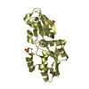
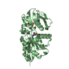
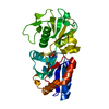
| Title | Crystal structure of a pheromone cOB1 precursor/lipoprotein, YaeC family (EF2496) from Enterococcus faecalis V583 at 2.10 A resolution | ||||||
 Components Components | Pheromone cOB1/lipoprotein, YaeC family | ||||||
 Keywords Keywords | METHIONINE-BINDING PROTEIN / periplasmic methionine binding protein / NLPA lipoprotein /  Structural Genomics / Joint Center for Structural Genomics / JCSG / Protein Structure Initiative / PSI-BIOLOGY Structural Genomics / Joint Center for Structural Genomics / JCSG / Protein Structure Initiative / PSI-BIOLOGY | ||||||
| Function / homology |  Function and homology information Function and homology information | ||||||
| Biological species |   Enterococcus faecalis (bacteria) Enterococcus faecalis (bacteria) | ||||||
| Method | X-RAY DIFFRACTION / SYNCHROTRON / MAD / Resolution: 2.1 Å | ||||||
 Authors Authors | Joint Center for Structural Genomics (JCSG) | ||||||
 Citation Citation | Journal: To be published Title: Crystal structure of a pheromone cOB1 precursor/lipoprotein, YaeC family (EF2496) from Enterococcus faecalis V583 at 2.10 A resolution Authors: Joint Center for Structural Genomics (JCSG) | ||||||
| History |
|
- Structure visualization
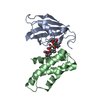
| Structure viewer | Molecule: MolmilJmol/JSmol |
|---|
- Downloads & links
-Download
| PDBx/mmCIF format | 4ef2.cif.gz | 202.1 KB | Display | PDBx/mmCIF format |
|---|---|---|---|---|
| PDB format | pdb4ef2.ent.gz | 165.1 KB | Display | PDB format |
| PDBx/mmJSON format | 4ef2.json.gz | Tree view | PDBx/mmJSON format | |
| Others |  Other downloads Other downloads |
-Validation report
| Arichive directory | https://data.pdbj.org/pub/pdb/validation_reports/ef/4ef2ftp://data.pdbj.org/pub/pdb/validation_reports/ef/4ef2 | HTTPS FTP |
|---|
-Related structure data
| Similar structure data | |
|---|---|
| Other databases |

-Links
 PDBj
PDBj


- Assembly
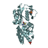
| Deposited unit | 
| ||||||||
|---|---|---|---|---|---|---|---|---|---|
| 1 | 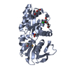
| ||||||||
| 2 | 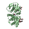
| ||||||||
| Unit cell |
|
-Components
-Protein , 1 types, 2 molecules AB
| #1: Protein | Mass: 27470.631 Da / Num. of mol.: 2 Source method: isolated from a genetically manipulated source Source: (gene. exp.) Enterococcus faecalis (bacteria) / Strain: V583 / Gene: EF_2496 / Plasmid: SpeedET / Production host: Escherichia Coli (E. coli) / Strain (production host): PB1 / References: UniProt: Q831K8 |
|---|
-Non-polymers , 6 types, 105 molecules 


| #2: Chemical | Selenomethionine Type: L-peptide linking / Mass: 196.106 Da / Num. of mol.: 2 / Source method: obtained synthetically / Formula: C5H11NO2Se Type: L-peptide linking / Mass: 196.106 Da / Num. of mol.: 2 / Source method: obtained synthetically / Formula: C5H11NO2Se#3: Chemical | ChemComp-CL / | Chloride Mass: 35.453 Da / Num. of mol.: 1 / Source method: obtained synthetically / Formula: Cl Mass: 35.453 Da / Num. of mol.: 1 / Source method: obtained synthetically / Formula: Cl#4: Chemical | ChemComp-PGE / | Polyethylene glycol Mass: 150.173 Da / Num. of mol.: 1 / Source method: obtained synthetically / Formula: C6H14O4 Mass: 150.173 Da / Num. of mol.: 1 / Source method: obtained synthetically / Formula: C6H14O4#5: Chemical | Diethylene glycol Mass: 106.120 Da / Num. of mol.: 2 / Source method: obtained synthetically / Formula: C4H10O3 Mass: 106.120 Da / Num. of mol.: 2 / Source method: obtained synthetically / Formula: C4H10O3#6: Chemical | ChemComp-PE8 / | Polyethylene glycol Mass: 370.436 Da / Num. of mol.: 1 / Source method: obtained synthetically / Formula: C16H34O9 Mass: 370.436 Da / Num. of mol.: 1 / Source method: obtained synthetically / Formula: C16H34O9#7: Water | ChemComp-HOH / | WaterMass: 18.015 Da / Num. of mol.: 98 / Source method: isolated from a natural source / Formula: H2O |
|---|
-Details
| Sequence details | THIS CONSTRUCT (RESIDUES 28-272) WAS EXPRESSED WITH A PURIFICATION TAG MGSDKIHHHHHHENLYFQG. THE TAG ...THIS CONSTRUCT (RESIDUES 28-272) WAS EXPRESSED WITH A PURIFICATI |
|---|
-Experimental details
-Experiment
| Experiment | Method: X-RAY DIFFRACTION / Number of used crystals: 1 |
|---|
- Sample preparation
| Crystal | Density Matthews: 2.29 Å3/Da / Density % sol: 46.28 % |
|---|---|
| Crystal grow | Temperature: 293 K / Method: vapor diffusion, sitting drop / pH: 9.5 Details: 30.0% polyethylene glycol 400, 0.1M CHES pH 9.5, NANODROP, VAPOR DIFFUSION, SITTING DROP, temperature 293K |
-Data collection
| Diffraction | Mean temperature: 100 K | |||||||||||||||||||||||||||||||||||||||||||||||||||||||||||||||||||||||||||||
|---|---|---|---|---|---|---|---|---|---|---|---|---|---|---|---|---|---|---|---|---|---|---|---|---|---|---|---|---|---|---|---|---|---|---|---|---|---|---|---|---|---|---|---|---|---|---|---|---|---|---|---|---|---|---|---|---|---|---|---|---|---|---|---|---|---|---|---|---|---|---|---|---|---|---|---|---|---|---|
| Diffraction source | Source: SYNCHROTRON / Site: SSRL  / Beamline: BL11-1 / Wavelength: 0.91837,0.97966,0.97901 / Beamline: BL11-1 / Wavelength: 0.91837,0.97966,0.97901 | |||||||||||||||||||||||||||||||||||||||||||||||||||||||||||||||||||||||||||||
| Detector | Type: DECTRIS PILATUS 6M / Detector: PIXEL / Date: Feb 8, 2012 Details: Flat mirror (vertical focusing); single crystal Si(111) bent monochromator (horizontal focusing) | |||||||||||||||||||||||||||||||||||||||||||||||||||||||||||||||||||||||||||||
| Radiation | Monochromator: single crystal Si(111) bent / Protocol: MAD / Monochromatic (M) / Laue (L): M / Scattering type: x-ray | |||||||||||||||||||||||||||||||||||||||||||||||||||||||||||||||||||||||||||||
| Radiation wavelength |
| |||||||||||||||||||||||||||||||||||||||||||||||||||||||||||||||||||||||||||||
| Reflection | Resolution: 2.1→28.763 Å / Num. obs: 28845 / % possible obs: 90.6 % / Observed criterion σ(I): -3 / Biso Wilson estimate: 38.876 Å2 / Rmerge(I) obs: 0.04 / Net I/σ(I): 12.37 | |||||||||||||||||||||||||||||||||||||||||||||||||||||||||||||||||||||||||||||
| Reflection shell | Diffraction-ID: 1
|
-Phasing
| Phasing | Method: MAD |
|---|
- Processing
| Software |
| ||||||||||||||||||||||||||||||||||||||||||||||||||||||||||||||||||||||||||||||||||||||||||
|---|---|---|---|---|---|---|---|---|---|---|---|---|---|---|---|---|---|---|---|---|---|---|---|---|---|---|---|---|---|---|---|---|---|---|---|---|---|---|---|---|---|---|---|---|---|---|---|---|---|---|---|---|---|---|---|---|---|---|---|---|---|---|---|---|---|---|---|---|---|---|---|---|---|---|---|---|---|---|---|---|---|---|---|---|---|---|---|---|---|---|---|
| Refinement | Method to determine structure: MAD / Resolution: 2.1→28.763 Å / Cor.coef. Fo:Fc: 0.961 / Cor.coef. Fo:Fc free: 0.943 / Occupancy max: 1 / Occupancy min: 0.5 / SU B: 11.455 / SU ML: 0.157 / Cross valid method: THROUGHOUT / σ(F): 0 / ESU R: 0.237 / ESU R Free: 0.191 Stereochemistry target values: MAXIMUM LIKELIHOOD WITH PHASES Details: 1.HYDROGENS HAVE BEEN ADDED IN THE RIDING POSITIONS. 2.A MET-INHIBITION PROTOCOL WAS USED FOR SELENOMETHIONINE INCORPORATION DURING PROTEIN EXPRESSION. THE OCCUPANCY OF THE SE ATOMS IN THE ...Details: 1.HYDROGENS HAVE BEEN ADDED IN THE RIDING POSITIONS. 2.A MET-INHIBITION PROTOCOL WAS USED FOR SELENOMETHIONINE INCORPORATION DURING PROTEIN EXPRESSION. THE OCCUPANCY OF THE SE ATOMS IN THE MSE RESIDUES WAS REDUCED TO 0.75 FOR THE REDUCED SCATTERING POWER DUE TO PARTIAL S-MET INCORPORATION. 3.ATOM RECORD CONTAINS SUM OF TLS AND RESIDUAL B FACTORS. ANISOU RECORD CONTAINS SUM OF TLS AND RESIDUAL U FACTORS. 4.WATERS WERE EXCLUDED FROM AUTOMATIC TLS ASSIGNMENT. 5.PEG FRAGMENTS (PEG, PGE AND PE8) FROM THE CRYSTALLIZATION SOLUTION AND CHLORIDE (CL) FROM THE PURIFICATION BUFFER HAVE BEEN MODELED IN THE SOLVENT STRUCTURE. 6.SELENO-METHIONINE (MSE) HAS BEEN MODELED AT THE PUTATIVE ACTIVE SITE BASED ON PUTATIVE ASSIGNMENT OF FUNCTION FROM PROTEIN STRUCTURE COMPARISONS AND ANOMALOUS DIFFERENCE FOURIER ELECTRON DENSITY MAPS. 7.RAMACHANDRAN OUTLIERS AT PHE84 IN BOTH PROTEIN ARE SUPPORTED BY ELECTRON DENSITY AND LIE IN THE VICINITY OF THE PUTATIVE ACTIVE SITE. 8.NCS RESTRAINTS WERE APPLIED USING REFMAC's LOCAL NCS OPTION (NCSR LOCAL). NCS GROUP 1 CHAIN A (30-270 TO CHAIN B (30-270) COUNT: 8183, RMS: 0.12, WEIGHT: 0.05.
| ||||||||||||||||||||||||||||||||||||||||||||||||||||||||||||||||||||||||||||||||||||||||||
| Solvent computation | Ion probe radii: 0.8 Å / Shrinkage radii: 0.8 Å / VDW probe radii: 1.2 Å / Solvent model: BABINET MODEL WITH MASK | ||||||||||||||||||||||||||||||||||||||||||||||||||||||||||||||||||||||||||||||||||||||||||
| Displacement parameters | Biso max: 121.37 Å2 / Biso mean: 52.5789 Å2 / Biso min: 23.5 Å2
| ||||||||||||||||||||||||||||||||||||||||||||||||||||||||||||||||||||||||||||||||||||||||||
| Refinement step | Cycle: LAST / Resolution: 2.1→28.763 Å
| ||||||||||||||||||||||||||||||||||||||||||||||||||||||||||||||||||||||||||||||||||||||||||
| Refine LS restraints |
| ||||||||||||||||||||||||||||||||||||||||||||||||||||||||||||||||||||||||||||||||||||||||||
| LS refinement shell | Resolution: 2.102→2.156 Å / Total num. of bins used: 20
| ||||||||||||||||||||||||||||||||||||||||||||||||||||||||||||||||||||||||||||||||||||||||||
| Refinement TLS params. | Method: refined / Refine-ID: X-RAY DIFFRACTION
| ||||||||||||||||||||||||||||||||||||||||||||||||||||||||||||||||||||||||||||||||||||||||||
| Refinement TLS group |
|
