 Movie
Movie Controller

[English] 日本語
 Yorodumi
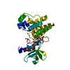
- PDB-3va4: Crystal structure of the mammalian MDC1 FHA domain complexed with... -
+ Open data

- Basic information
| Entry | Database: PDB / ID: 3va4 | ||||||
|---|---|---|---|---|---|---|---|
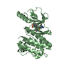
| Title | Crystal structure of the mammalian MDC1 FHA domain complexed with CHK2 pThr68 peptide | ||||||
 Components Components |
| ||||||
 Keywords Keywords |  CELL CYCLE / FHA domain / DNA-damage / CHK2 and MDC1 dimerization CELL CYCLE / FHA domain / DNA-damage / CHK2 and MDC1 dimerization | ||||||
| Function / homology |  Function and homology information Function and homology informationSUMOylation of DNA damage response and repair proteins / G2/M DNA damage checkpoint / Nonhomologous End-Joining (NHEJ) / Processing of DNA double-strand break ends / mitotic DNA damage checkpoint signaling / Recruitment and ATM-mediated phosphorylation of repair and signaling proteins at DNA double strand breaks / protein localization to site of double-strand break / DNA replication checkpoint signaling / mitotic intra-S DNA damage checkpoint signaling / DNA damage response, signal transduction by p53 class mediator resulting in transcription of p21 class mediator ...SUMOylation of DNA damage response and repair proteins / G2/M DNA damage checkpoint / Nonhomologous End-Joining (NHEJ) / Processing of DNA double-strand break ends / mitotic DNA damage checkpoint signaling / Recruitment and ATM-mediated phosphorylation of repair and signaling proteins at DNA double strand breaks / protein localization to site of double-strand break / DNA replication checkpoint signaling / mitotic intra-S DNA damage checkpoint signaling / DNA damage response, signal transduction by p53 class mediator resulting in transcription of p21 class mediator / thymocyte apoptotic process / regulation of protein catabolic process / replicative senescence / mitotic spindle assembly / positive regulation of transcription initiation by RNA polymerase II / intrinsic apoptotic signaling pathway in response to DNA damage by p53 class mediator / signal transduction in response to DNA damage / Chk1/Chk2(Cds1) mediated inactivation of Cyclin B:Cdk1 complex / DNA damage response, signal transduction by p53 class mediator resulting in cell cycle arrest / histone reader activity / regulation of signal transduction by p53 class mediator / DNA damage checkpoint signaling / Ubiquitin Mediated Degradation of Phosphorylated Cdc25A / Stabilization of p53 / protein catabolic process / G2/M DNA damage checkpoint / Regulation of TP53 Activity through Methylation / cellular response to gamma radiation / PML body / G2/M transition of mitotic cell cycle / intrinsic apoptotic signaling pathway in response to DNA damage / double-strand break repair / Regulation of TP53 Degradation / Recruitment and ATM-mediated phosphorylation of repair and signaling proteins at DNA double strand breaks / site of double-strand break / chromosome / Regulation of TP53 Activity through Phosphorylation / protein autophosphorylation / nuclear body / protein stabilization / non-specific serine/threonine protein kinase / cell cycle / cell division / protein phosphorylation / protein serine kinase activity / DNA repair / focal adhesion / protein serine/threonine kinase activity / DNA damage response / ubiquitin protein ligase binding / regulation of DNA-templated transcription / protein kinase binding / positive regulation of DNA-templated transcription / Golgi apparatus / protein homodimerization activity / nucleoplasm / ATP binding / identical protein binding / metal ion binding / nucleus / cytoplasmSimilarity search - Function | ||||||
| Biological species |  Mus musculus (house mouse) Mus musculus (house mouse) Homo sapiens (human) Homo sapiens (human) | ||||||
| Method | X-RAY DIFFRACTION / SYNCHROTRON / SAD / Resolution: 1.54 Å | ||||||
 Authors Authors | Wu, H.H. / Wu, P.Y. / Huang, K.F. / Kao, Y.Y. / Tsai, M.D. | ||||||
 Citation Citation | Journal: Biochemistry / Year: 2012 Title: Structural Delineation of MDC1-FHA Domain Binding with CHK2-pThr68. Authors: Wu, H.H. / Wu, P.Y. / Huang, K.F. / Kao, Y.Y. / Tsai, M.D. | ||||||
| History |
|
- Structure visualization
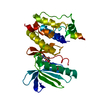
| Structure viewer | Molecule: MolmilJmol/JSmol |
|---|
- Downloads & links
-Download
| PDBx/mmCIF format | 3va4.cif.gz | 104.5 KB | Display | PDBx/mmCIF format |
|---|---|---|---|---|
| PDB format | pdb3va4.ent.gz | 86.2 KB | Display | PDB format |
| PDBx/mmJSON format | 3va4.json.gz | Tree view | PDBx/mmJSON format | |
| Others |  Other downloads Other downloads |
-Validation report
| Arichive directory | https://data.pdbj.org/pub/pdb/validation_reports/va/3va4ftp://data.pdbj.org/pub/pdb/validation_reports/va/3va4 | HTTPS FTP |
|---|
-Related structure data


-Links
 PDBj
PDBj


- Assembly
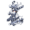
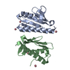
| Deposited unit | 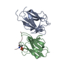
| ||||||||
|---|---|---|---|---|---|---|---|---|---|
| 1 |
| ||||||||
| Unit cell |
|
-Components
| #1: Protein | Mass: 14589.714 Da / Num. of mol.: 2 / Fragment: N-terminal FHA domain (UNP RESIDUES 29-139) Source method: isolated from a genetically manipulated source Source: (gene. exp.) Mus musculus (house mouse) / Strain: Mouse / Gene: MDC1 / Plasmid: pET28a / Production host:  Escherichia coli (E. coli) / Strain (production host): BL21 / References: UniProt: Q5PSV9 Escherichia coli (E. coli) / Strain (production host): BL21 / References: UniProt: Q5PSV9#2: Protein/peptide | | Mass: 1349.335 Da / Num. of mol.: 1 / Fragment: CHK2-pThr68 peptide (UNP RESIDUES 63-73) / Source method: obtained synthetically / Details: The peptide was chemically synthesized. / Source: (synth.) Homo sapiens (human) / References: UniProt: O96017#3: Water | ChemComp-HOH / | Water Mass: 18.015 Da / Num. of mol.: 153 / Source method: isolated from a natural source / Formula: H2O Mass: 18.015 Da / Num. of mol.: 153 / Source method: isolated from a natural source / Formula: H2O |
|---|
-Experimental details
-Experiment
| Experiment | Method: X-RAY DIFFRACTION / Number of used crystals: 1 |
|---|
- Sample preparation
| Crystal | Density % sol: 25.96 % |
|---|---|
| Crystal grow | Temperature: 293 K / Method: vapor diffusion, sitting drop / pH: 9.5 Details: 1.26M (NH4)2SO4, 0.2M NaCl, 0.1M CHES, pH 9.5, VAPOR DIFFUSION, SITTING DROP, temperature 293K |
-Data collection
| Diffraction | Mean temperature: 100 K |
|---|---|
| Diffraction source | Source: SYNCHROTRON / Site: NSRRC  / Beamline: BL13B1 / Wavelength: 0.9762 Å / Beamline: BL13B1 / Wavelength: 0.9762 Å |
| Detector | Type: ADSC QUANTUM 315r / Detector: CCD / Date: Mar 5, 2011 |
| Radiation | Monochromator: GRAPHITE / Protocol: SINGLE WAVELENGTH / Monochromatic (M) / Laue (L): M / Scattering type: x-ray |
| Radiation wavelength | Wavelength: 0.9762 Å / Relative weight: 1 |
| Reflection | Resolution: 1.54→57.72 Å / Num. all: 31096 / Num. obs: 30878 / % possible obs: 99.3 % / Observed criterion σ(F): 2 / Observed criterion σ(I): 3 / Redundancy: 4.6 % / Rmerge(I) obs: 0.037 / Rsym value: 0.037 / Net I/σ(I): 31.5 |
| Reflection shell | Resolution: 1.54→1.6 Å / Redundancy: 4.5 % / Rmerge(I) obs: 0.508 / Mean I/σ(I) obs: 2.2 / Num. unique all: 3039 / Rsym value: 0.508 / % possible all: 99.3 |
- Processing
| Software |
| |||||||||||||||||||||||||||||||||||||||||||||||||||||||||||||||||||||||||||
|---|---|---|---|---|---|---|---|---|---|---|---|---|---|---|---|---|---|---|---|---|---|---|---|---|---|---|---|---|---|---|---|---|---|---|---|---|---|---|---|---|---|---|---|---|---|---|---|---|---|---|---|---|---|---|---|---|---|---|---|---|---|---|---|---|---|---|---|---|---|---|---|---|---|---|---|---|
| Refinement | Method to determine structure: SAD / Resolution: 1.54→50 Å / Cor.coef. Fo:Fc: 0.969 / Cor.coef. Fo:Fc free: 0.958 / SU B: 3.104 / SU ML: 0.051 / Cross valid method: THROUGHOUT / ESU R: 0.102 / ESU R Free: 0.083 / Stereochemistry target values: MAXIMUM LIKELIHOOD / Details: HYDROGENS HAVE BEEN USED IF PRESENT IN THE INPUT
| |||||||||||||||||||||||||||||||||||||||||||||||||||||||||||||||||||||||||||
| Solvent computation | Ion probe radii: 0.8 Å / Shrinkage radii: 0.8 Å / VDW probe radii: 1.2 Å / Solvent model: MASK | |||||||||||||||||||||||||||||||||||||||||||||||||||||||||||||||||||||||||||
| Displacement parameters | Biso mean: 23.648 Å2
| |||||||||||||||||||||||||||||||||||||||||||||||||||||||||||||||||||||||||||
| Refinement step | Cycle: LAST / Resolution: 1.54→50 Å
| |||||||||||||||||||||||||||||||||||||||||||||||||||||||||||||||||||||||||||
| Refine LS restraints |
| |||||||||||||||||||||||||||||||||||||||||||||||||||||||||||||||||||||||||||
| LS refinement shell | Resolution: 1.54→1.58 Å / Total num. of bins used: 20
| |||||||||||||||||||||||||||||||||||||||||||||||||||||||||||||||||||||||||||
| Refinement TLS params. | Method: refined / Refine-ID: X-RAY DIFFRACTION
| |||||||||||||||||||||||||||||||||||||||||||||||||||||||||||||||||||||||||||
| Refinement TLS group |
|
