 Movie
Movie Controller


+ Open data

- Basic information
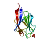
| Entry | Database: PDB / ID: 3qx1 | ||||||
|---|---|---|---|---|---|---|---|
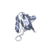
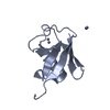
| Title | Crystal structure of FAF1 UBX domain | ||||||
 Components Components | FAS-associated factor 1 | ||||||
 Keywords Keywords |  PROTEIN BINDING / UBX / p97 Binding PROTEIN BINDING / UBX / p97 Binding | ||||||
| Function / homology |  Function and homology informationooplasm / positive regulation of extrinsic apoptotic signaling pathway via death domain receptors / CD95 death-inducing signaling complex / cytoplasmic sequestering of NF-kappaB / protein kinase regulator activity / VCP-NPL4-UFD1 AAA ATPase complex / regulation of protein catabolic process / : / NF-kappaB binding / regulation of cell adhesion ...ooplasm / positive regulation of extrinsic apoptotic signaling pathway via death domain receptors / CD95 death-inducing signaling complex / cytoplasmic sequestering of NF-kappaB / protein kinase regulator activity / VCP-NPL4-UFD1 AAA ATPase complex / regulation of protein catabolic process / : / NF-kappaB binding / regulation of cell adhesion / heat shock protein binding / ubiquitin binding / positive regulation of DNA replication / positive regulation of protein-containing complex assembly / positive regulation of protein catabolic process / nuclear envelope / proteasome-mediated ubiquitin-dependent protein catabolic process / positive regulation of apoptotic process / protein domain specific binding / apoptotic process / ubiquitin protein ligase binding / protein kinase binding / perinuclear region of cytoplasm / endoplasmic reticulum / nucleoplasm / nucleus / cytosol Function and homology informationooplasm / positive regulation of extrinsic apoptotic signaling pathway via death domain receptors / CD95 death-inducing signaling complex / cytoplasmic sequestering of NF-kappaB / protein kinase regulator activity / VCP-NPL4-UFD1 AAA ATPase complex / regulation of protein catabolic process / : / NF-kappaB binding / regulation of cell adhesion ...ooplasm / positive regulation of extrinsic apoptotic signaling pathway via death domain receptors / CD95 death-inducing signaling complex / cytoplasmic sequestering of NF-kappaB / protein kinase regulator activity / VCP-NPL4-UFD1 AAA ATPase complex / regulation of protein catabolic process / : / NF-kappaB binding / regulation of cell adhesion / heat shock protein binding / ubiquitin binding / positive regulation of DNA replication / positive regulation of protein-containing complex assembly / positive regulation of protein catabolic process / nuclear envelope / proteasome-mediated ubiquitin-dependent protein catabolic process / positive regulation of apoptotic process / protein domain specific binding / apoptotic process / ubiquitin protein ligase binding / protein kinase binding / perinuclear region of cytoplasm / endoplasmic reticulum / nucleoplasm / nucleus / cytosolSimilarity search - Function | ||||||
| Biological species |  Homo sapiens (human) Homo sapiens (human) | ||||||
| Method | X-RAY DIFFRACTION / SYNCHROTRON / MOLECULAR REPLACEMENT / Resolution: 1.6 Å | ||||||
 Authors Authors | Park, J.K. / Jeon, H. / Lee, J.J. / Kim, K.H. / Lee, K.J. / Kim, E.E. | ||||||
 Citation Citation | Journal: To be Published Title: Dissection of the interaction between FAF1 UBX and p97 Authors: Park, J.K. / Jeon, H. / Lee, J.J. / Kim, K.H. / Lee, K.J. / Kim, E.E. | ||||||
| History |
|
- Structure visualization
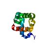
| Structure viewer | Molecule: MolmilJmol/JSmol |
|---|
- Downloads & links
-Download
| PDBx/mmCIF format | 3qx1.cif.gz | 52.2 KB | Display | PDBx/mmCIF format |
|---|---|---|---|---|
| PDB format | pdb3qx1.ent.gz | 36.9 KB | Display | PDB format |
| PDBx/mmJSON format | 3qx1.json.gz | Tree view | PDBx/mmJSON format | |
| Others |  Other downloads Other downloads |
-Validation report
| Arichive directory | https://data.pdbj.org/pub/pdb/validation_reports/qx/3qx1ftp://data.pdbj.org/pub/pdb/validation_reports/qx/3qx1 | HTTPS FTP |
|---|
-Related structure data
| Related structure data |  3qwzSC S: Starting model for refinement C: citing same article ( |
|---|---|
| Similar structure data |


-Links
 PDBj
PDBj- Assembly
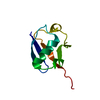
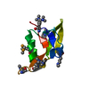
| Deposited unit | 
| ||||||||
|---|---|---|---|---|---|---|---|---|---|
| 1 | 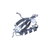
| ||||||||
| 2 | 
| ||||||||
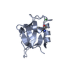
| Unit cell |
| ||||||||
| Components on special symmetry positions |
|
-Components
| #1: Protein | Mass: 9828.306 Da / Num. of mol.: 2 / Fragment: FAF1 UBX domain, UNP residues 571-650 Source method: isolated from a genetically manipulated source Source: (gene. exp.) Homo sapiens (human) / Gene: CGI-03, FAF1, UBXD12, UBXN3A / Plasmid: pET28a / Production host:  Escherichia coli (E. coli) / Strain (production host): E.coli Rosetta (DE3) / References: UniProt: Q9UNN5 Escherichia coli (E. coli) / Strain (production host): E.coli Rosetta (DE3) / References: UniProt: Q9UNN5#2: Chemical | Sulfate  Mass: 96.063 Da / Num. of mol.: 2 / Source method: obtained synthetically / Formula: SO4 Mass: 96.063 Da / Num. of mol.: 2 / Source method: obtained synthetically / Formula: SO4#3: Water | ChemComp-HOH / | Water Mass: 18.015 Da / Num. of mol.: 228 / Source method: isolated from a natural source / Formula: H2O Mass: 18.015 Da / Num. of mol.: 228 / Source method: isolated from a natural source / Formula: H2O |
|---|
-Experimental details
-Experiment
| Experiment | Method: X-RAY DIFFRACTION / Number of used crystals: 1 |
|---|
- Sample preparation
| Crystal | Density Matthews: 2.09 Å3/Da / Density % sol: 41.2 % |
|---|---|
| Crystal grow | Temperature: 295 K / Method: vapor diffusion, hanging drop / pH: 7.5 Details: 30% PEG 3350, 0.2M Lithium sulfate, 0.1M HEPES pH7.5, VAPOR DIFFUSION, HANGING DROP, temperature 295K |
-Data collection
| Diffraction | Mean temperature: 100 K |
|---|---|
| Diffraction source | Source: SYNCHROTRON / Site: PAL/PLS  / Beamline: 4A / Wavelength: 1 Å / Beamline: 4A / Wavelength: 1 Å |
| Detector | Type: ADSC QUANTUM 315 / Detector: CCD / Date: Oct 17, 2007 |
| Radiation | Protocol: SINGLE WAVELENGTH / Monochromatic (M) / Laue (L): M / Scattering type: x-ray |
| Radiation wavelength | Wavelength: 1 Å / Relative weight: 1 |
| Reflection | Resolution: 1.6→20 Å / Num. obs: 21633 / % possible obs: 98.4 % / Observed criterion σ(F): 1 / Observed criterion σ(I): 1 / Biso Wilson estimate: 18.5 Å2 / Rmerge(I) obs: 0.079 |
| Reflection shell | Resolution: 1.6→1.66 Å / Rmerge(I) obs: 0.276 / Mean I/σ(I) obs: 5.4 / % possible all: 96.4 |
- Processing
| Software |
| ||||||||||||||||||||||||||||||||||||||||||||||||||||||||||||||||||||||||||||||||
|---|---|---|---|---|---|---|---|---|---|---|---|---|---|---|---|---|---|---|---|---|---|---|---|---|---|---|---|---|---|---|---|---|---|---|---|---|---|---|---|---|---|---|---|---|---|---|---|---|---|---|---|---|---|---|---|---|---|---|---|---|---|---|---|---|---|---|---|---|---|---|---|---|---|---|---|---|---|---|---|---|---|
| Refinement | Method to determine structure: MOLECULAR REPLACEMENT Starting model: PDB ENTRY 3QWZ Resolution: 1.6→19.95 Å / Rfactor Rfree error: 0.007 / Data cutoff high absF: 119062.48 / Data cutoff low absF: 0 / Isotropic thermal model: RESTRAINED / Cross valid method: THROUGHOUT / σ(F): 0 / Stereochemistry target values: ENGH & HUBER
| ||||||||||||||||||||||||||||||||||||||||||||||||||||||||||||||||||||||||||||||||
| Solvent computation | Solvent model: FLAT MODEL / Bsol: 45.42 Å2 / ksol: 0.38 e/Å3 | ||||||||||||||||||||||||||||||||||||||||||||||||||||||||||||||||||||||||||||||||
| Displacement parameters | Biso mean: 22.4 Å2
| ||||||||||||||||||||||||||||||||||||||||||||||||||||||||||||||||||||||||||||||||
| Refine analyze |
| ||||||||||||||||||||||||||||||||||||||||||||||||||||||||||||||||||||||||||||||||
| Refinement step | Cycle: LAST / Resolution: 1.6→19.95 Å
| ||||||||||||||||||||||||||||||||||||||||||||||||||||||||||||||||||||||||||||||||
| Refine LS restraints |
| ||||||||||||||||||||||||||||||||||||||||||||||||||||||||||||||||||||||||||||||||
| Refine LS restraints NCS | NCS model details: NONE | ||||||||||||||||||||||||||||||||||||||||||||||||||||||||||||||||||||||||||||||||
| LS refinement shell | Resolution: 1.6→1.7 Å / Rfactor Rfree error: 0.021 / Total num. of bins used: 6
| ||||||||||||||||||||||||||||||||||||||||||||||||||||||||||||||||||||||||||||||||
| Xplor file |
|
