 Movie
Movie Controller


+ Open data

- Basic information
| Entry | Database: PDB / ID: 2zpm | ||||||
|---|---|---|---|---|---|---|---|
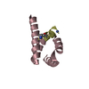
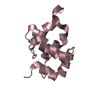
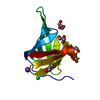
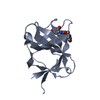
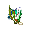
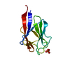
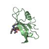
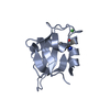
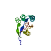
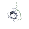
| Title | Crystal structure analysis of PDZ domain B | ||||||
 Components Components | Regulator of sigma E protease | ||||||
 Keywords Keywords |  HYDROLASE / metalloproteinase / membrane protein / PDZ domain / Inner membrane / Membrane / Metal-binding / Metalloprotease / Protease / Transmembrane / Zinc HYDROLASE / metalloproteinase / membrane protein / PDZ domain / Inner membrane / Membrane / Metal-binding / Metalloprotease / Protease / Transmembrane / Zinc | ||||||
| Function / homology |  Function and homology information Function and homology informationanti-sigma factor antagonist activity / Hydrolases; Acting on peptide bonds (peptidases); Metalloendopeptidases / cellular response to cell envelope stress / metalloendopeptidase activity / positive regulation of DNA-templated transcription / proteolysis / metal ion binding / plasma membraneSimilarity search - Function | ||||||
| Biological species |  Escherichia coli (E. coli) Escherichia coli (E. coli) | ||||||
| Method | X-RAY DIFFRACTION / SYNCHROTRON / SAD / Resolution: 0.98 Å | ||||||
 Authors Authors | Inaba, K. / Suzuki, M. | ||||||
 Citation Citation | Journal: To be Published Title: Crystal Structure analysis of PDZ-domain B Authors: Inaba, K. / Suzuki, M. / Maegawa, K. / Akiyama, Y. | ||||||
| History |
|
- Structure visualization
| Structure viewer | Molecule: MolmilJmol/JSmol |
|---|
- Downloads & links
-Download
| PDBx/mmCIF format | 2zpm.cif.gz | 50.8 KB | Display | PDBx/mmCIF format |
|---|---|---|---|---|
| PDB format | pdb2zpm.ent.gz | 41.5 KB | Display | PDB format |
| PDBx/mmJSON format | 2zpm.json.gz | Tree view | PDBx/mmJSON format | |
| Others |  Other downloads Other downloads |
-Validation report
| Arichive directory | https://data.pdbj.org/pub/pdb/validation_reports/zp/2zpmftp://data.pdbj.org/pub/pdb/validation_reports/zp/2zpm | HTTPS FTP |
|---|
-Related structure data
| Similar structure data |
|---|
-Links
 PDBj
PDBj
- Assembly
| Deposited unit | 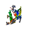
| ||||||||||||
|---|---|---|---|---|---|---|---|---|---|---|---|---|---|
| 1 |
| ||||||||||||
| 2 | 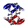
| ||||||||||||
| Unit cell |
| ||||||||||||
| Components on special symmetry positions |
|
-Components
| #1: Protein | Mass: 9815.257 Da / Num. of mol.: 1 / Fragment: PDZ-domain B Source method: isolated from a genetically manipulated source Source: (gene. exp.) Escherichia coli (E. coli) / Strain: K12 / Plasmid: pGEX-5X-1 / Production host: Escherichia coli (E. coli)References: UniProt: P0AEH1, Hydrolases; Acting on peptide bonds (peptidases); Metalloendopeptidases |
|---|---|
| #2: Water | ChemComp-HOH / Water Mass: 18.015 Da / Num. of mol.: 151 / Source method: isolated from a natural source / Formula: H2O Mass: 18.015 Da / Num. of mol.: 151 / Source method: isolated from a natural source / Formula: H2O |
-Experimental details
-Experiment
| Experiment | Method: X-RAY DIFFRACTION / Number of used crystals: 1 |
|---|
- Sample preparation
| Crystal | Density Matthews: 1.9 Å3/Da / Density % sol: 35.35 % |
|---|---|
| Crystal grow | Temperature: 293 K / Method: vapor diffusion / Details: 25% PEG3350, VAPOR DIFFUSION, temperature 293K |
-Data collection
| Diffraction | Mean temperature: 100 K |
|---|---|
| Diffraction source | Source: SYNCHROTRON / Site: SPring-8  / Beamline: BL44XU / Wavelength: 0.65 Å / Beamline: BL44XU / Wavelength: 0.65 Å |
| Detector | Type: Bruker DIP-6040 / Detector: CCD |
| Radiation | Protocol: SINGLE WAVELENGTH / Monochromatic (M) / Laue (L): M / Scattering type: x-ray |
| Radiation wavelength | Wavelength: 0.65 Å / Relative weight: 1 |
| Reflection | Resolution: 0.98→21.47 Å / Num. obs: 42333 / % possible obs: 99.4 % / Redundancy: 3.7 % / Biso Wilson estimate: 7.3 Å2 / Rmerge(I) obs: 0.044 / Net I/σ(I): 17.1 |
| Reflection shell | Resolution: 0.98→1.03 Å / Redundancy: 3.7 % / Rmerge(I) obs: 0.446 / Mean I/σ(I) obs: 2.9 / % possible all: 99 |
- Processing
| Software |
| ||||||||||||||||||||||||||||||||||||||||||||||||||||||||||||||||||||||||||||||||
|---|---|---|---|---|---|---|---|---|---|---|---|---|---|---|---|---|---|---|---|---|---|---|---|---|---|---|---|---|---|---|---|---|---|---|---|---|---|---|---|---|---|---|---|---|---|---|---|---|---|---|---|---|---|---|---|---|---|---|---|---|---|---|---|---|---|---|---|---|---|---|---|---|---|---|---|---|---|---|---|---|---|
| Refinement | Method to determine structure: SAD / Resolution: 0.98→20.75 Å / Cor.coef. Fo:Fc: 0.974 / Cor.coef. Fo:Fc free: 0.964 / Cross valid method: THROUGHOUT / ESU R: 0.025 / ESU R Free: 0.027 / Stereochemistry target values: MAXIMUM LIKELIHOOD
| ||||||||||||||||||||||||||||||||||||||||||||||||||||||||||||||||||||||||||||||||
| Solvent computation | Ion probe radii: 0.8 Å / Shrinkage radii: 0.8 Å / VDW probe radii: 1.4 Å / Solvent model: BABINET MODEL WITH MASK | ||||||||||||||||||||||||||||||||||||||||||||||||||||||||||||||||||||||||||||||||
| Displacement parameters | Biso mean: 13.227 Å2
| ||||||||||||||||||||||||||||||||||||||||||||||||||||||||||||||||||||||||||||||||
| Refinement step | Cycle: LAST / Resolution: 0.98→20.75 Å
| ||||||||||||||||||||||||||||||||||||||||||||||||||||||||||||||||||||||||||||||||
| Refine LS restraints |
| ||||||||||||||||||||||||||||||||||||||||||||||||||||||||||||||||||||||||||||||||
| LS refinement shell | Resolution: 0.981→1.006 Å / Total num. of bins used: 20
|
