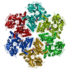
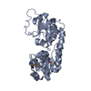
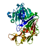
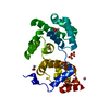
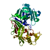
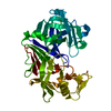
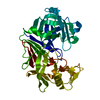
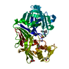
Entry Database : PDB / ID : 3qwzTitle Crystal structure of FAF1 UBX-p97N-domain complex FAS-associated factor 1 Transitional endoplasmic reticulum ATPase Keywords / / Function / homology Function Domain/homology Component
/ / / / / / / / / / / / / / / / / / / / / / / / / / / / / / / / / / / / / / / / / / / / / / / / / / / / / / / / / / / / / / / / / / / / / / / / / / / / / / / / / / / / / / / / / / / / / / / / / / / / / / / / / / / / / / / / / / / / / / / / / / / / / / / / / / / / / / / / / / / / / / / / / / / / / / / / / / / / / Biological species Homo sapiens (human)Method / / / Resolution : 2 Å Authors Park, J.K. / Jeon, H. / Lee, J.J. / Kim, K.H. / Lee, K.J. / Kim, E.E. Journal : To be Published Title : Dissection of the interaction between FAF1 UBX and p97Authors : Park, J.K. / Jeon, H. / Lee, J.J. / Kim, K.H. / Lee, K.J. / Kim, E.E. History Deposition Feb 28, 2011 Deposition site / Processing site Revision 1.0 May 9, 2012 Provider / Type Revision 1.1 Sep 13, 2023 Group Data collection / Database references ... Data collection / Database references / Derived calculations / Refinement description Category chem_comp_atom / chem_comp_bond ... chem_comp_atom / chem_comp_bond / database_2 / pdbx_initial_refinement_model / struct_conn / struct_ref_seq_dif Item _database_2.pdbx_DOI / _database_2.pdbx_database_accession ... _database_2.pdbx_DOI / _database_2.pdbx_database_accession / _struct_conn.pdbx_leaving_atom_flag / _struct_ref_seq_dif.details
Show all Show less
 Movie
Movie Controller


 Open data
 Basic information
 Components
 Keywords
 TRANSPORT PROTEIN / UBX / p97 Binding
 Function and homology information
 Authors
 Citation
 Structure visualization
 Downloads & links
 Other downloads
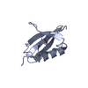


 PDBj
PDBj


 Assembly
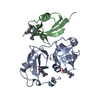


 Mass: 18.015 Da / Num. of mol.: 163 / Source method: isolated from a natural source / Formula: H2O
 Sample preparation
 / Beamline: 4A / Wavelength: 1
  Processing