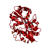
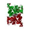
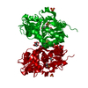
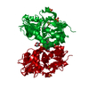
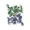
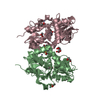
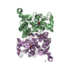
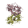
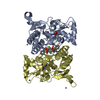
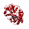
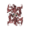
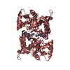
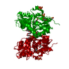
Mass: 29177.670 Da / Num. of mol.: 1 Fragment: Ligand binding domain, residues 413 to 527 and 653 to 796 Source method: isolated from a genetically manipulated source Details: S1-S2 fusion in which Gly118 and Thr119 replace a membrane-spanning region Source: (gene. exp.) Rattus norvegicus (Norway rat) / Gene: Gria2, Glur2 / Plasmid: pET-32a / Production host: Escherichia coli (E. coli) / Strain (production host): Origami B (de3) / References: UniProt: P19491
Monochromator: Graphite / Protocol: SINGLE WAVELENGTH / Monochromatic (M) / Laue (L): M / Scattering type: x-ray
Radiation wavelength
Wavelength: 1.54178 Å / Relative weight: 1
Reflection
Resolution: 1.87→26.73 Å / Num. all: 21208 / Num. obs: 21208 / % possible obs: 92.9 % / Observed criterion σ(I): 2 / Redundancy: 4.4 % / Biso Wilson estimate: 31.2 Å2 / Rsym value: 0.051 / Net I/σ(I): 17.3
Reflection shell
Resolution: 1.87→1.92 Å / Redundancy: 4 % / Rmerge(I) obs: 0.661 / Mean I/σ(I) obs: 1.9 / Num. unique all: 1442 / % possible all: 86.5
-
Processing
Software
Name
Version
Classification
CrystalClear
datacollection
AMoRE
phasing
REFMAC
5.2.0019
refinement
MOSFLM
datareduction
SCALA
datascaling
Refinement
Method to determine structure: MOLECULAR REPLACEMENT / Resolution: 1.87→26.73 Å / Cor.coef. Fo:Fc: 0.952 / Cor.coef. Fo:Fc free: 0.923 / SU B: 5.501 / SU ML: 0.162 / Cross valid method: THROUGHOUT / ESU R Free: 0.19 / Stereochemistry target values: MAXIMUM LIKELIHOOD / Details: HYDROGENS HAVE BEEN ADDED IN THE RIDING POSITIONS
Rfactor
Num. reflection
% reflection
Selection details
Rfree
0.28142
1089
5.1 %
RANDOM
Rwork
0.21754
-
-
-
all
0.22086
20080
-
-
obs
0.22086
20080
100 %
-
Solvent computation
Ion probe radii: 0.8 Å / Shrinkage radii: 0.8 Å / VDW probe radii: 1.4 Å / Solvent model: MASK
Displacement parameters
Biso mean: 21.358 Å2
Baniso -1
Baniso -2
Baniso -3
1-
-2.95 Å2
0 Å2
0 Å2
2-
-
3.07 Å2
0 Å2
3-
-
-
-0.12 Å2
Refinement step
Cycle: LAST / Resolution: 1.87→26.73 Å
Protein
Nucleic acid
Ligand
Solvent
Total
Num. atoms
2043
0
92
311
2446
Refine LS restraints
Refine-ID
Type
Dev ideal
X-RAY DIFFRACTION
r_bond_refined_d
0.014
X-RAY DIFFRACTION
r_bond_other_d
0.006
X-RAY DIFFRACTION
r_angle_refined_deg
1.442
X-RAY DIFFRACTION
r_angle_other_deg
0.976
X-RAY DIFFRACTION
r_dihedral_angle_1_deg
5.999
X-RAY DIFFRACTION
r_dihedral_angle_2_deg
30.736
X-RAY DIFFRACTION
r_dihedral_angle_3_deg
15.804
X-RAY DIFFRACTION
r_dihedral_angle_4_deg
21.885
X-RAY DIFFRACTION
r_chiral_restr
0.078
X-RAY DIFFRACTION
r_gen_planes_refined
0.005
X-RAY DIFFRACTION
r_gen_planes_other
0.001
X-RAY DIFFRACTION
r_nbd_refined
0.199
X-RAY DIFFRACTION
r_nbd_other
0.207
X-RAY DIFFRACTION
r_nbtor_refined
0.17
X-RAY DIFFRACTION
r_nbtor_other
0.087
X-RAY DIFFRACTION
r_xyhbond_nbd_refined
0.172
X-RAY DIFFRACTION
r_symmetry_vdw_refined
0.159
X-RAY DIFFRACTION
r_symmetry_vdw_other
0.28
X-RAY DIFFRACTION
r_symmetry_hbond_refined
0.247
X-RAY DIFFRACTION
r_mcbond_it
1.492
X-RAY DIFFRACTION
r_mcbond_other
0.258
X-RAY DIFFRACTION
r_mcangle_it
1.599
X-RAY DIFFRACTION
r_scbond_it
2.545
X-RAY DIFFRACTION
r_scangle_it
3.277
LS refinement shell
Resolution: 1.87→1.916 Å / Total num. of bins used: 20
Rfactor
Num. reflection
% reflection
Rfree
0.517
87
-
Rwork
0.411
1347
-
obs
-
-
100 %
+
About Yorodumi
-
News
-
Feb 9, 2022. New format data for meta-information of EMDB entries
New format data for meta-information of EMDB entries
Version 3 of the EMDB header file is now the official format.
The previous official version 1.9 will be removed from the archive.
In the structure databanks used in Yorodumi, some data are registered as the other names, "COVID-19 virus" and "2019-nCoV". Here are the details of the virus and the list of structure data.
Jan 31, 2019. EMDB accession codes are about to change! (news from PDBe EMDB page)
EMDB accession codes are about to change! (news from PDBe EMDB page)
The allocation of 4 digits for EMDB accession codes will soon come to an end. Whilst these codes will remain in use, new EMDB accession codes will include an additional digit and will expand incrementally as the available range of codes is exhausted. The current 4-digit format prefixed with “EMD-” (i.e. EMD-XXXX) will advance to a 5-digit format (i.e. EMD-XXXXX), and so on. It is currently estimated that the 4-digit codes will be depleted around Spring 2019, at which point the 5-digit format will come into force.
The EM Navigator/Yorodumi systems omit the EMD- prefix.
Related info.:Q: What is EMD? / ID/Accession-code notation in Yorodumi/EM Navigator
Yorodumi is a browser for structure data from EMDB, PDB, SASBDB, etc.
This page is also the successor to EM Navigator detail page, and also detail information page/front-end page for Omokage search.
The word "yorodu" (or yorozu) is an old Japanese word meaning "ten thousand". "mi" (miru) is to see.
Related info.:EMDB / PDB / SASBDB / Comparison of 3 databanks / Yorodumi Search / Aug 31, 2016. New EM Navigator & Yorodumi / Yorodumi Papers / Jmol/JSmol / Function and homology information / Changes in new EM Navigator and Yorodumi
 Movie
Movie Controller

 Yorodumi
 Open data
 Basic information
 Components
 GRIA2
GRIA2  Keywords
 Function and homology information
 Authors
 Citation
 Structure visualization
 Downloads & links
 Other downloads


 PDBj
PDBj


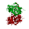
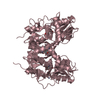
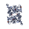
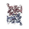
 Assembly


 Type: L-peptide linking / Mass: 147.129 Da / Num. of mol.: 1 / Source method: obtained synthetically / Formula: C5H9NO4
 Mass: 96.063 Da / Num. of mol.: 4 / Source method: obtained synthetically / Formula: SO4
 Mass: 92.094 Da / Num. of mol.: 2 / Source method: obtained synthetically / Formula: C3H8O3
 Mass: 78.133 Da / Num. of mol.: 2 / Source method: obtained synthetically / Formula: C2H6OS / Comment: DMSO, precipitant*YM
 Mass: 62.068 Da / Num. of mol.: 4 / Source method: obtained synthetically / Formula: C2H6O2
 Mass: 387.417 Da / Num. of mol.: 1 / Source method: obtained synthetically / Formula: C18H20F3NO3S
 Sample preparation
 Processing