Mass: 18.015 Da / Num. of mol.: 389 / Source method: isolated from a natural source / Formula: H2O
-
Details
Sequence details
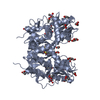
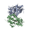
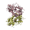
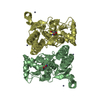
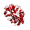
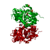
THE SEQUENCE MATCHES UNIPROT ENTRY P19491 ISOFORM FLIP WITH IDENTIFIER P19491-2. IT IS AN S1-S2 ...THE SEQUENCE MATCHES UNIPROT ENTRY P19491 ISOFORM FLIP WITH IDENTIFIER P19491-2. IT IS AN S1-S2 FUSION PROTEIN IN WHICH G118 AND T119 REPLACE RESIDUES 528-652 OF THE NATIVE PROTEIN.
-
Experimental details
-
Experiment
Experiment
Method: X-RAY DIFFRACTION / Number of used crystals: 1
-
Sample preparation
Crystal
Density Matthews: 2.29 Å3/Da / Density % sol: 46.19 %
Crystal grow
Temperature: 298 K / Method: vapor diffusion, hanging drop / pH: 4.1 Details: 18% PEG 4000, 50mM Lithium Sulphate, 100mM Sodium Cacodylate, pH 4.1, VAPOR DIFFUSION, HANGING DROP, temperature 298K
Resolution: 1.4→25 Å / Cor.coef. Fo:Fc: 0.956 / Cor.coef. Fo:Fc free: 0.935 / SU B: 0.92 / SU ML: 0.038 / Cross valid method: THROUGHOUT / ESU R: 0.068 / ESU R Free: 0.074 / Stereochemistry target values: MAXIMUM LIKELIHOOD / Details: HYDROGENS HAVE BEEN ADDED IN THE RIDING POSITIONS
Rfactor
Num. reflection
% reflection
Selection details
Rfree
0.20938
2495
5.2 %
RANDOM
Rwork
0.16808
-
-
-
all
0.188
48434
-
-
obs
0.17016
45898
90.9 %
-
Solvent computation
Ion probe radii: 0.8 Å / Shrinkage radii: 0.8 Å / VDW probe radii: 1.2 Å / Solvent model: BABINET MODEL WITH MASK
Displacement parameters
Biso mean: 14.912 Å2
Baniso -1
Baniso -2
Baniso -3
1-
-0.6 Å2
0 Å2
0 Å2
2-
-
1 Å2
0 Å2
3-
-
-
-0.4 Å2
Refinement step
Cycle: LAST / Resolution: 1.4→25 Å
Protein
Nucleic acid
Ligand
Solvent
Total
Num. atoms
2043
0
77
389
2509
Refine LS restraints
Refine-ID
Type
Dev ideal
Dev ideal target
Number
X-RAY DIFFRACTION
r_bond_refined_d
0.021
0.02
2328
X-RAY DIFFRACTION
r_bond_other_d
0.002
0.02
1651
X-RAY DIFFRACTION
r_angle_refined_deg
2.291
2.009
3144
X-RAY DIFFRACTION
r_angle_other_deg
2.901
3.001
4079
X-RAY DIFFRACTION
r_dihedral_angle_1_deg
6.173
5
295
X-RAY DIFFRACTION
r_dihedral_angle_2_deg
29.224
24.194
93
X-RAY DIFFRACTION
r_dihedral_angle_3_deg
14.972
15
467
X-RAY DIFFRACTION
r_dihedral_angle_4_deg
24.731
15
12
X-RAY DIFFRACTION
r_chiral_restr
0.154
0.2
342
X-RAY DIFFRACTION
r_gen_planes_refined
0.011
0.02
2506
X-RAY DIFFRACTION
r_gen_planes_other
0.001
0.02
451
LS refinement shell
Resolution: 1.4→1.438 Å / Total num. of bins used: 20
Rfactor
Num. reflection
% reflection
Rfree
0.213
110
-
Rwork
0.189
2143
-
obs
-
2203
59.13 %
+
About Yorodumi
-
News
-
Feb 9, 2022. New format data for meta-information of EMDB entries
New format data for meta-information of EMDB entries
Version 3 of the EMDB header file is now the official format.
The previous official version 1.9 will be removed from the archive.
In the structure databanks used in Yorodumi, some data are registered as the other names, "COVID-19 virus" and "2019-nCoV". Here are the details of the virus and the list of structure data.
Jan 31, 2019. EMDB accession codes are about to change! (news from PDBe EMDB page)
EMDB accession codes are about to change! (news from PDBe EMDB page)
The allocation of 4 digits for EMDB accession codes will soon come to an end. Whilst these codes will remain in use, new EMDB accession codes will include an additional digit and will expand incrementally as the available range of codes is exhausted. The current 4-digit format prefixed with “EMD-” (i.e. EMD-XXXX) will advance to a 5-digit format (i.e. EMD-XXXXX), and so on. It is currently estimated that the 4-digit codes will be depleted around Spring 2019, at which point the 5-digit format will come into force.
The EM Navigator/Yorodumi systems omit the EMD- prefix.
Related info.:Q: What is EMD? / ID/Accession-code notation in Yorodumi/EM Navigator
Yorodumi is a browser for structure data from EMDB, PDB, SASBDB, etc.
This page is also the successor to EM Navigator detail page, and also detail information page/front-end page for Omokage search.
The word "yorodu" (or yorozu) is an old Japanese word meaning "ten thousand". "mi" (miru) is to see.
Related info.:EMDB / PDB / SASBDB / Comparison of 3 databanks / Yorodumi Search / Aug 31, 2016. New EM Navigator & Yorodumi / Yorodumi Papers / Jmol/JSmol / Function and homology information / Changes in new EM Navigator and Yorodumi
 Movie
Movie Controller

 Yorodumi
 Open data
 Basic information
 Components
 GRIA2
GRIA2  Keywords
 Function and homology information
 Authors
 Citation
 Structure visualization
 Downloads & links
 Other downloads
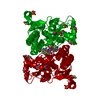


 PDBj
PDBj


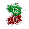
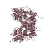
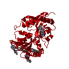
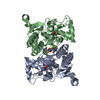
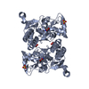
 Assembly


 Type: L-peptide linking / Mass: 147.129 Da / Num. of mol.: 1 / Source method: obtained synthetically / Formula: C5H9NO4
 Mass: 402.391 Da / Num. of mol.: 1 / Source method: obtained synthetically / Formula: C16H17F3N4O3S
 Mass: 96.063 Da / Num. of mol.: 3 / Source method: obtained synthetically / Formula: SO4
 Mass: 92.094 Da / Num. of mol.: 4 / Source method: obtained synthetically / Formula: C3H8O3
 Mass: 6.941 Da / Num. of mol.: 1 / Source method: obtained synthetically / Formula: Li
 Sample preparation
 / Beamline: ID23-1 / Wavelength: 0.95375 Å
 Processing