 ムービー
ムービー コントローラー


+ データを開く

- 基本情報
基本情報
| 登録情報 | データベース: PDB / ID: 1mqd | ||||||
|---|---|---|---|---|---|---|---|
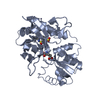
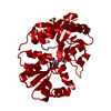
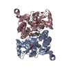
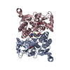
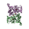
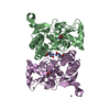
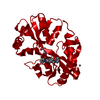
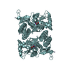
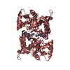
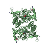
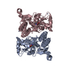
| タイトル | X-ray structure of the GluR2 ligand-binding core (S1S2J) in complex with (S)-Des-Me-AMPA at 1.46 A resolution. Crystallization in the presence of lithium sulfate. | ||||||
 要素 要素 | Glutamate receptor subunit 2 グルタミン酸受容体 グルタミン酸受容体 | ||||||
 キーワード キーワード | MEMBRANE PROTEIN (膜タンパク質) / Ionotropic glutamate receptor GluR2 / ligand-binding core / agonist complex. (アゴニスト) | ||||||
| 機能・相同性 |  機能・相同性情報 機能・相同性情報spine synapse / dendritic spine neck / dendritic spine head / Activation of AMPA receptors / response to lithium ion / perisynaptic space / cellular response to glycine / AMPA glutamate receptor activity / Trafficking of GluR2-containing AMPA receptors / immunoglobulin binding ...spine synapse / dendritic spine neck / dendritic spine head / Activation of AMPA receptors / response to lithium ion / perisynaptic space / cellular response to glycine / AMPA glutamate receptor activity / Trafficking of GluR2-containing AMPA receptors / immunoglobulin binding / AMPA glutamate receptor complex / kainate selective glutamate receptor activity / ionotropic glutamate receptor complex / extracellularly glutamate-gated ion channel activity / asymmetric synapse / regulation of receptor recycling / Unblocking of NMDA receptors, glutamate binding and activation / glutamate receptor binding / positive regulation of synaptic transmission / glutamate-gated receptor activity / presynaptic active zone membrane / response to fungicide / regulation of synaptic transmission, glutamatergic / cellular response to brain-derived neurotrophic factor stimulus / somatodendritic compartment / dendrite membrane / ligand-gated monoatomic ion channel activity involved in regulation of presynaptic membrane potential / ionotropic glutamate receptor binding / ionotropic glutamate receptor signaling pathway / dendrite cytoplasm / cytoskeletal protein binding / SNARE binding / transmitter-gated monoatomic ion channel activity involved in regulation of postsynaptic membrane potential / dendritic shaft / synaptic membrane / synaptic transmission, glutamatergic / PDZ domain binding / postsynaptic density membrane / protein tetramerization / modulation of chemical synaptic transmission / Schaffer collateral - CA1 synapse / establishment of protein localization / terminal bouton / receptor internalization / synaptic vesicle membrane / cerebral cortex development / シナプス小胞 / presynapse / signaling receptor activity / presynaptic membrane / amyloid-beta binding / 成長円錐 / perikaryon / chemical synaptic transmission / scaffold protein binding / postsynaptic membrane / 樹状突起スパイン / postsynaptic density / neuron projection / 神経繊維 / neuronal cell body / 樹状突起 / シナプス / glutamatergic synapse / protein-containing complex binding / endoplasmic reticulum membrane / protein kinase binding / 細胞膜 / 小胞体 / protein-containing complex / 生体膜 / identical protein binding / 細胞膜類似検索 - 分子機能 | ||||||
| 生物種 |  Rattus norvegicus (ドブネズミ) Rattus norvegicus (ドブネズミ) | ||||||
| 手法 | X線回折 / シンクロトロン / 分子置換 / 解像度: 1.46 Å | ||||||
 データ登録者 データ登録者 | Kasper, C. / Lunn, M.-L. / Liljefors, T. / Gouaux, E. / Egebjerg, J. / Kastrup, J.S. | ||||||
 引用 引用 | ジャーナル: FEBS Lett. / 年: 2002 タイトル: GluR2 ligand-binding core complexes: Importance of the isoxazolol moiety and 5-substituent for the binding mode of AMPA-type agonists. 著者: Kasper, C. / Lunn, M.L. / Liljefors, T. / Gouaux, E. / Egebjerg, J. / Kastrup, J.S. #1: ジャーナル: J.Mol.Biol. / 年: 2002タイトル: Structural basis for AMPA receptor activation and ligand selectivity: Crystal structures of five agonist complexes with the GluR2 ligand binding core. 著者: Hogner, A. / Kastrup, J. / Jin, R. / Liljefors, T. / Mayer, M. / Egebjerg, J. / Larsen, I. / Gouaux, E. #2: ジャーナル: Neuron / 年: 2000タイトル: Mechanisms for activation and antagonism of an AMPA-sensitive glutamate receptor: Crystal structures of the GluR2 ligand binding core. 著者: Armstrong, N. / Gouaux, E. #3: ジャーナル: Nature / 年: 2002タイトル: Mechanism of glutamate receptor desensitization. 著者: Sun, Y. / Olson, R. / Horning, M. / Armstrong, N. / Mayer, M. / Gouaux, E. #4: ジャーナル: Protein Sci. / 年: 1998タイトル: Probing the ligand binding domain of the GluR2 receptor by proteolysis and deletion mutagenesis defines domain boundaries and yields a crystallizable construct. 著者: Chen, G.Q. / Sun, Y. / Jin, R. / Gouaux, E. | ||||||
| 履歴 |
|
- 構造の表示
構造の表示
| 構造ビューア | 分子: MolmilJmol/JSmol |
|---|
- ダウンロードとリンク
-ダウンロード
| PDBx/mmCIF形式 | 1mqd.cif.gz | 279.3 KB | 表示 | PDBx/mmCIF形式 |
|---|---|---|---|---|
| PDB形式 | pdb1mqd.ent.gz | 220 KB | 表示 | PDB形式 |
| PDBx/mmJSON形式 | 1mqd.json.gz | ツリー表示 | PDBx/mmJSON形式 | |
| その他 |  その他のダウンロード その他のダウンロード |
-検証レポート
| アーカイブディレクトリ | https://data.pdbj.org/pub/pdb/validation_reports/mq/1mqdftp://data.pdbj.org/pub/pdb/validation_reports/mq/1mqd | HTTPS FTP |
|---|
-関連構造データ


-リンク
 PDBj
PDBj


- 集合体
集合体
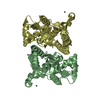
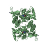
| 登録構造単位 | 
| ||||||||||||
|---|---|---|---|---|---|---|---|---|---|---|---|---|---|
| 1 | 
| ||||||||||||
| 2 | 
| ||||||||||||
| 3 | 
| ||||||||||||
| 4 | 
| ||||||||||||
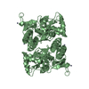
| 単位格子 |
| ||||||||||||
| Components on special symmetry positions |
| ||||||||||||
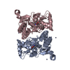
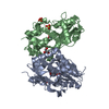
| 詳細 | The biological assembly is a dimer. The dimer of chain A can be generated by -x, y, -z, shift 1 0 0. The dimer of chain B can be generated by -x, y, -z, shift 0 0 0. The dimer of chain C can be generated by -x, y, -z, shift -1 0 -1. The dimer of chain D can be generated by -x, y, -z, shift 0 0 -1. |
-要素
| #1: タンパク質 | グルタミン酸受容体 / GluR-2 分子量: 29077.553 Da / 分子数: 4 / 断片: GluR2-flop ligand-binding core (S1S2J) / 由来タイプ: 組換発現 / 詳細: Tetherd dimer linked by GLY 115 and THR 116 / 由来: (組換発現) Rattus norvegicus (ドブネズミ) / プラスミド: pET30B / 生物種 (発現宿主): Escherichia coli / 発現宿主:  Escherichia coli BL21(DE3) (大腸菌) / 株 (発現宿主): BL21 (DE3) / 参照: UniProt: P19491 Escherichia coli BL21(DE3) (大腸菌) / 株 (発現宿主): BL21 (DE3) / 参照: UniProt: P19491#2: 化合物 | 硫酸塩  分子量: 96.063 Da / 分子数: 3 / 由来タイプ: 合成 / 式: SO4 分子量: 96.063 Da / 分子数: 3 / 由来タイプ: 合成 / 式: SO4#3: 化合物 | ChemComp-SHI / (   分子量: 172.139 Da / 分子数: 4 / 由来タイプ: 合成 / 式: C6H8N2O4 分子量: 172.139 Da / 分子数: 4 / 由来タイプ: 合成 / 式: C6H8N2O4#4: 化合物 | ChemComp-GOL / | グリセリン  分子量: 92.094 Da / 分子数: 1 / 由来タイプ: 合成 / 式: C3H8O3 分子量: 92.094 Da / 分子数: 1 / 由来タイプ: 合成 / 式: C3H8O3#5: 水 | ChemComp-HOH / | 水 分子量: 18.015 Da / 分子数: 2626 / 由来タイプ: 天然 / 式: H2O 分子量: 18.015 Da / 分子数: 2626 / 由来タイプ: 天然 / 式: H2O |
|---|
-実験情報
-実験
| 実験 | 手法: X線回折 / 使用した結晶の数: 1 |
|---|
- 試料調製
試料調製
| 結晶 | マシュー密度: 2.44 Å3/Da / 溶媒含有率: 49.65 % | |||||||||||||||||||||||||||||||||||||||||||||||||||||||||||||||
|---|---|---|---|---|---|---|---|---|---|---|---|---|---|---|---|---|---|---|---|---|---|---|---|---|---|---|---|---|---|---|---|---|---|---|---|---|---|---|---|---|---|---|---|---|---|---|---|---|---|---|---|---|---|---|---|---|---|---|---|---|---|---|---|---|
| 結晶化 | 温度: 279 K / 手法: 蒸気拡散法, ハンギングドロップ法 / pH: 5.2 詳細: 20% PEG 8000, 0.1M lithium sulfate, 0.1M cacodylate, pH 5.2, VAPOR DIFFUSION, HANGING DROP, temperature 279K | |||||||||||||||||||||||||||||||||||||||||||||||||||||||||||||||
| 結晶化 | *PLUS 温度: 6 ℃ / pH: 7.4 / 手法: 蒸気拡散法, ハンギングドロップ法 | |||||||||||||||||||||||||||||||||||||||||||||||||||||||||||||||
| 溶液の組成 | *PLUS
|
-データ収集
| 回折 | 平均測定温度: 100 K |
|---|---|
| 放射光源 | 由来: シンクロトロン / サイト: EMBL/DESY, HAMBURG  / ビームライン: X11 / 波長: 0.8499 Å / ビームライン: X11 / 波長: 0.8499 Å |
| 検出器 | タイプ: MARRESEARCH / 検出器: CCD / 日付: 2001年8月15日 |
| 放射 | プロトコル: SINGLE WAVELENGTH / 単色(M)・ラウエ(L): M / 散乱光タイプ: x-ray |
| 放射波長 | 波長: 0.8499 Å / 相対比: 1 |
| 反射 | 解像度: 1.46→20 Å / Num. all: 191888 / Num. obs: 191888 / % possible obs: 99 % / Observed criterion σ(F): 0 / Observed criterion σ(I): 0 / 冗長度: 3.9 % / Biso Wilson estimate: 14.9 Å2 / Rmerge(I) obs: 0.042 / Net I/σ(I): 26.7 |
| 反射 シェル | 解像度: 1.46→1.49 Å / Rmerge(I) obs: 0.384 / Mean I/σ(I) obs: 2.4 / Num. unique all: 12223 / % possible all: 95.5 |
| 反射 | *PLUS 最低解像度: 20 Å |
| 反射 シェル | *PLUS % possible obs: 95.5 % |
- 解析
解析
| ソフトウェア |
| |||||||||||||||||||||||||
|---|---|---|---|---|---|---|---|---|---|---|---|---|---|---|---|---|---|---|---|---|---|---|---|---|---|---|
| 精密化 | 構造決定の手法: 分子置換 開始モデル: PDB entry 1M5B 解像度: 1.46→19.24 Å / Rfactor Rfree error: 0.003 / Data cutoff high absF: 1705947 / Data cutoff high rms absF: 1705947 / Isotropic thermal model: RESTRAINED / 交差検証法: THROUGHOUT / σ(F): 0 / σ(I): 0 / 立体化学のターゲット値: Engh & Huber 詳細: The first three N-terminal residues and the last two C-terminal residues were not located in the electron density map. The side chains of the following residues are not fully defined: Lys A1, ...詳細: The first three N-terminal residues and the last two C-terminal residues were not located in the electron density map. The side chains of the following residues are not fully defined: Lys A1, Lys A18, Lys A66, Lys A104, Lys A126, Glu A129, Glu A142, Arg A146, Lys A180, Lys A246, Lys B1, Lys B18, Glu B21, Glu B24, Ala B63, Asp B64, Lys B66, Glu B119, Lys B126, Glu B142, Lys B255, Lys C1, Lys C47, Arg C61, Asp C64, Lys C66, Lys C126, Glu C129, Glu C142, Arg C146, Lys C148, Arg C180, Lys C246, Lys C255, Lys D1, Lys D18, Met D22, Lys D47, Lys D49, Ala D63, Lys D66, Lys D126, Glu D142
| |||||||||||||||||||||||||
| 溶媒の処理 | 溶媒モデル: FLAT MODEL / Bsol: 52.86 Å2 / ksol: 0.34 e/Å3 | |||||||||||||||||||||||||
| 原子変位パラメータ | Biso mean: 21.2 Å2
| |||||||||||||||||||||||||
| Refine analyze |
| |||||||||||||||||||||||||
| 精密化ステップ | サイクル: LAST / 解像度: 1.46→19.24 Å
| |||||||||||||||||||||||||
| 拘束条件 |
| |||||||||||||||||||||||||
| LS精密化 シェル | 解像度: 1.46→1.55 Å / Rfactor Rfree error: 0.011 / Total num. of bins used: 6
| |||||||||||||||||||||||||
| Xplor file |
| |||||||||||||||||||||||||
| 精密化 | *PLUS 最低解像度: 20 Å / % reflection Rfree: 2 % / Rfactor Rwork: 0.18 | |||||||||||||||||||||||||
| 溶媒の処理 | *PLUS | |||||||||||||||||||||||||
| 原子変位パラメータ | *PLUS | |||||||||||||||||||||||||
| 拘束条件 | *PLUS
|
