 Movie
Movie Controller


+ Open data

- Basic information
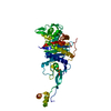
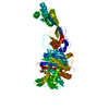
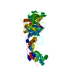
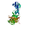
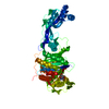
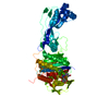
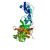
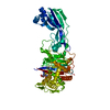
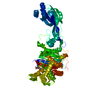
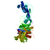
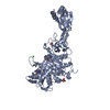
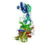
| Entry | Database: PDB / ID: 3pbt | ||||||
|---|---|---|---|---|---|---|---|
| Title | Crystal structure of PBP3 complexed with MC-1 | ||||||
 Components Components | Penicillin-binding protein 3 | ||||||
 Keywords Keywords | HYDROLASE/ANTIBIOTIC / PBP3 / HYDROLASE-ANTIBIOTIC complex | ||||||
| Function / homology |  Function and homology information Function and homology information peptidoglycan glycosyltransferase activity / serine-type D-Ala-D-Ala carboxypeptidase / FtsZ-dependent cytokinesis / serine-type D-Ala-D-Ala carboxypeptidase activity / division septum assembly / penicillin binding / peptidoglycan biosynthetic process / cell wall organization / regulation of cell shape / proteolysis / plasma membrane peptidoglycan glycosyltransferase activity / serine-type D-Ala-D-Ala carboxypeptidase / FtsZ-dependent cytokinesis / serine-type D-Ala-D-Ala carboxypeptidase activity / division septum assembly / penicillin binding / peptidoglycan biosynthetic process / cell wall organization / regulation of cell shape / proteolysis / plasma membraneSimilarity search - Function | ||||||
| Biological species |   Pseudomonas aeruginosa (bacteria) Pseudomonas aeruginosa (bacteria) | ||||||
| Method | X-RAY DIFFRACTION / SYNCHROTRON / MOLECULAR REPLACEMENT / Resolution: 1.641 Å | ||||||
 Authors Authors | Han, S. / Evdokimov, A. | ||||||
 Citation Citation | Journal: Proc.Natl.Acad.Sci.USA / Year: 2010 Title: Structural basis for effectiveness of siderophore-conjugated monocarbams against clinically relevant strains of Pseudomonas aeruginosa. Authors: Han, S. / Zaniewski, R.P. / Marr, E.S. / Lacey, B.M. / Tomaras, A.P. / Evdokimov, A. / Miller, J.R. / Shanmugasundaram, V. | ||||||
| History |
|
- Structure visualization
| Structure viewer | Molecule: MolmilJmol/JSmol |
|---|
- Downloads & links
-Download
| PDBx/mmCIF format | 3pbt.cif.gz | 121.5 KB | Display | PDBx/mmCIF format |
|---|---|---|---|---|
| PDB format | pdb3pbt.ent.gz | 98.8 KB | Display | PDB format |
| PDBx/mmJSON format | 3pbt.json.gz | Tree view | PDBx/mmJSON format | |
| Others |  Other downloads Other downloads |
-Validation report
| Arichive directory | https://data.pdbj.org/pub/pdb/validation_reports/pb/3pbtftp://data.pdbj.org/pub/pdb/validation_reports/pb/3pbt | HTTPS FTP |
|---|
-Related structure data


-Links
 PDBj
PDBj
- Assembly
| Deposited unit | 
| ||||||||
|---|---|---|---|---|---|---|---|---|---|
| 1 |
| ||||||||
| Unit cell |
|
-Components
| #1: Protein | Mass: 58329.453 Da / Num. of mol.: 1 / Fragment: UNP Residues 50-579 Source method: isolated from a genetically manipulated source Source: (gene. exp.) Pseudomonas aeruginosa (bacteria) / Strain: PAO1 / Gene: ftsI, PA4418, pbpB / Production host: Escherichia coli (E. coli) / References: UniProt: Q51504, UniProt: G3XD46*PLUS |
|---|---|
| #2: Chemical | ChemComp-UE1 / (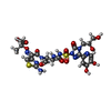  Mass: 715.650 Da / Num. of mol.: 1 / Source method: obtained synthetically / Formula: C23H27N10O13S2 Mass: 715.650 Da / Num. of mol.: 1 / Source method: obtained synthetically / Formula: C23H27N10O13S2 |
| #3: Water | ChemComp-HOH / Water Mass: 18.015 Da / Num. of mol.: 539 / Source method: isolated from a natural source / Formula: H2O Mass: 18.015 Da / Num. of mol.: 539 / Source method: isolated from a natural source / Formula: H2O |
-Experimental details
-Experiment
| Experiment | Method: X-RAY DIFFRACTION / Number of used crystals: 1 |
|---|
- Sample preparation
| Crystal | Density Matthews: 2.18 Å3/Da / Density % sol: 43.56 % |
|---|---|
| Crystal grow | Method: vapor diffusion, hanging drop / pH: 6.2 Details: 0.1M citrate, 30% PEG3350, 0.3M Mg(NO3)2, pH 6.2, VAPOR DIFFUSION, HANGING DROP |
-Data collection
| Diffraction | Mean temperature: 100 K |
|---|---|
| Diffraction source | Source: SYNCHROTRON / Site: APS  / Beamline: 17-ID / Wavelength: 1 Å / Beamline: 17-ID / Wavelength: 1 Å |
| Detector | Type: ADSC QUANTUM 210 / Detector: CCD / Date: Aug 1, 2009 |
| Radiation | Protocol: SINGLE WAVELENGTH / Monochromatic (M) / Laue (L): M / Scattering type: x-ray |
| Radiation wavelength | Wavelength: 1 Å / Relative weight: 1 |
| Reflection | Resolution: 1.64→61.12 Å / Num. obs: 62097 / % possible obs: 99.1 % / Observed criterion σ(F): 1 / Observed criterion σ(I): 1 |
- Processing
| Software |
| ||||||||||||||||||||||||||||||||||||||||||||||||||||||||||||||||||||||||||||||||||||||||||||||||||||||||||||||||||||||||||||||||||||||||||||||||||||||||||||||||||||||||||
|---|---|---|---|---|---|---|---|---|---|---|---|---|---|---|---|---|---|---|---|---|---|---|---|---|---|---|---|---|---|---|---|---|---|---|---|---|---|---|---|---|---|---|---|---|---|---|---|---|---|---|---|---|---|---|---|---|---|---|---|---|---|---|---|---|---|---|---|---|---|---|---|---|---|---|---|---|---|---|---|---|---|---|---|---|---|---|---|---|---|---|---|---|---|---|---|---|---|---|---|---|---|---|---|---|---|---|---|---|---|---|---|---|---|---|---|---|---|---|---|---|---|---|---|---|---|---|---|---|---|---|---|---|---|---|---|---|---|---|---|---|---|---|---|---|---|---|---|---|---|---|---|---|---|---|---|---|---|---|---|---|---|---|---|---|---|---|---|---|---|---|---|
| Refinement | Method to determine structure: MOLECULAR REPLACEMENT / Resolution: 1.641→61.12 Å / Cor.coef. Fo:Fc: 0.965 / Cor.coef. Fo:Fc free: 0.95 / SU B: 1.788 / SU ML: 0.063 / Cross valid method: THROUGHOUT / ESU R: 0.097 / ESU R Free: 0.096 / Stereochemistry target values: MAXIMUM LIKELIHOOD / Details: HYDROGENS HAVE BEEN ADDED IN THE RIDING POSITIONS
| ||||||||||||||||||||||||||||||||||||||||||||||||||||||||||||||||||||||||||||||||||||||||||||||||||||||||||||||||||||||||||||||||||||||||||||||||||||||||||||||||||||||||||
| Solvent computation | Ion probe radii: 0.8 Å / Shrinkage radii: 0.8 Å / VDW probe radii: 1.2 Å / Solvent model: MASK | ||||||||||||||||||||||||||||||||||||||||||||||||||||||||||||||||||||||||||||||||||||||||||||||||||||||||||||||||||||||||||||||||||||||||||||||||||||||||||||||||||||||||||
| Displacement parameters | Biso mean: 22.645 Å2
| ||||||||||||||||||||||||||||||||||||||||||||||||||||||||||||||||||||||||||||||||||||||||||||||||||||||||||||||||||||||||||||||||||||||||||||||||||||||||||||||||||||||||||
| Refinement step | Cycle: LAST / Resolution: 1.641→61.12 Å
| ||||||||||||||||||||||||||||||||||||||||||||||||||||||||||||||||||||||||||||||||||||||||||||||||||||||||||||||||||||||||||||||||||||||||||||||||||||||||||||||||||||||||||
| Refine LS restraints |
| ||||||||||||||||||||||||||||||||||||||||||||||||||||||||||||||||||||||||||||||||||||||||||||||||||||||||||||||||||||||||||||||||||||||||||||||||||||||||||||||||||||||||||
| LS refinement shell | Resolution: 1.641→1.684 Å / Total num. of bins used: 20
|
