 Movie
Movie Controller

[English] 日本語
 Yorodumi
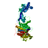
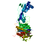
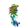
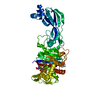
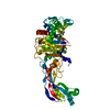
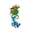
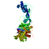
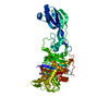
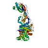
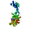
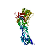
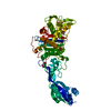
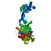
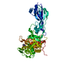
- PDB-3ocn: Crystal structure of penicillin-binding protein 3 from Pseudomona... -
+ Open data

- Basic information
| Entry | Database: PDB / ID: 3ocn | ||||||
|---|---|---|---|---|---|---|---|
| Title | Crystal structure of penicillin-binding protein 3 from Pseudomonas aeruginosa in complex with ceftazidime | ||||||
 Components Components | penicillin-binding protein 3 | ||||||
 Keywords Keywords | PENICILLIN-BINDING PROTEIN/ANTIBIOTIC /  penicillin-binding proteins / Pseudomonas aeruginosa / ceftazidime / Structural Genomics / Oxford Protein Production Facility / OPPF / transpeptidase / cell wall biosynthesis / out periplasmic membrane / PENICILLIN-BINDING PROTEIN-ANTIBIOTIC complex penicillin-binding proteins / Pseudomonas aeruginosa / ceftazidime / Structural Genomics / Oxford Protein Production Facility / OPPF / transpeptidase / cell wall biosynthesis / out periplasmic membrane / PENICILLIN-BINDING PROTEIN-ANTIBIOTIC complex | ||||||
| Function / homology |  Function and homology informationpeptidoglycan glycosyltransferase activity / serine-type D-Ala-D-Ala carboxypeptidase / FtsZ-dependent cytokinesis / serine-type D-Ala-D-Ala carboxypeptidase activity / division septum assembly / penicillin binding / peptidoglycan biosynthetic process / cell wall organization / regulation of cell shape / proteolysis / plasma membrane Function and homology informationpeptidoglycan glycosyltransferase activity / serine-type D-Ala-D-Ala carboxypeptidase / FtsZ-dependent cytokinesis / serine-type D-Ala-D-Ala carboxypeptidase activity / division septum assembly / penicillin binding / peptidoglycan biosynthetic process / cell wall organization / regulation of cell shape / proteolysis / plasma membraneSimilarity search - Function | ||||||
| Biological species |   Pseudomonas aeruginosa (bacteria) Pseudomonas aeruginosa (bacteria) | ||||||
| Method | X-RAY DIFFRACTION / SYNCHROTRON / MOLECULAR REPLACEMENT / Resolution: 2.61 Å | ||||||
 Authors Authors | Sainsbury, S. / Bird, L. / Stuart, D.I. / Owens, R.J. / Ren, J. / Oxford Protein Production Facility (OPPF) | ||||||
 Citation Citation | Journal: J.Mol.Biol. / Year: 2011 Title: Crystal structures of penicillin-binding protein 3 from Pseudomonas aeruginosa: comparison of native and antibiotic-bound forms Authors: Sainsbury, S. / Bird, L. / Rao, V. / Shepherd, S.M. / Stuart, D.I. / Hunter, W.N. / Owens, R.J. / Ren, J. | ||||||
| History |
|
- Structure visualization
| Structure viewer | Molecule: MolmilJmol/JSmol |
|---|
- Downloads & links
-Download
| PDBx/mmCIF format | 3ocn.cif.gz | 207 KB | Display | PDBx/mmCIF format |
|---|---|---|---|---|
| PDB format | pdb3ocn.ent.gz | 163 KB | Display | PDB format |
| PDBx/mmJSON format | 3ocn.json.gz | Tree view | PDBx/mmJSON format | |
| Others |  Other downloads Other downloads |
-Validation report
| Arichive directory | https://data.pdbj.org/pub/pdb/validation_reports/oc/3ocnftp://data.pdbj.org/pub/pdb/validation_reports/oc/3ocn | HTTPS FTP |
|---|
-Related structure data
| Related structure data | 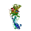 3oc2C  3oclSC C: citing same article ( S: Starting model for refinement |
|---|---|
| Similar structure data |


-Links
 PDBj
PDBj
- Assembly
| Deposited unit | 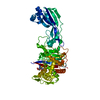
| ||||||||
|---|---|---|---|---|---|---|---|---|---|
| 1 |
| ||||||||
| Unit cell |
|
-Components
| #1: Protein | Mass: 61152.590 Da / Num. of mol.: 1 / Fragment: residues in UNP 35-579 Source method: isolated from a genetically manipulated source Source: (gene. exp.) Pseudomonas aeruginosa (bacteria) / Strain: PAO1 / Gene: PA4418 / Plasmid: OPPF6502 / Production host: Escherichia coli (E. coli) / Strain (production host): DE3 / References: UniProt: Q51504 |
|---|---|
| #2: Chemical | ChemComp-CTJ /   Mass: 549.600 Da / Num. of mol.: 1 / Source method: obtained synthetically / Formula: C22H25N6O7S2 Mass: 549.600 Da / Num. of mol.: 1 / Source method: obtained synthetically / Formula: C22H25N6O7S2 |
| #3: Water | ChemComp-HOH / Water Mass: 18.015 Da / Num. of mol.: 38 / Source method: isolated from a natural source / Formula: H2O Mass: 18.015 Da / Num. of mol.: 38 / Source method: isolated from a natural source / Formula: H2O |
-Experimental details
-Experiment
| Experiment | Method: X-RAY DIFFRACTION / Number of used crystals: 1 |
|---|
- Sample preparation
| Crystal | Density Matthews: 1.97 Å3/Da / Density % sol: 37.55 % |
|---|---|
| Crystal grow | Temperature: 294 K / Method: vapor diffusion, sitting drop / pH: 7.8 Details: 25%(w/v) polyethylene glycol 3350, 0.1M Bis-Tris propane, 1%(w/v) protamine sulphate, pH 7.8, VAPOR DIFFUSION, SITTING DROP, temperature 294K |
-Data collection
| Diffraction | Mean temperature: 100 K |
|---|---|
| Diffraction source | Source: SYNCHROTRON / Site: Diamond  / Beamline: I02 / Wavelength: 0.975 Å / Beamline: I02 / Wavelength: 0.975 Å |
| Detector | Type: ADSC QUANTUM 315 / Detector: CCD / Date: Mar 7, 2010 |
| Radiation | Protocol: SINGLE WAVELENGTH / Monochromatic (M) / Laue (L): M / Scattering type: x-ray |
| Radiation wavelength | Wavelength: 0.975 Å / Relative weight: 1 |
| Reflection | Resolution: 2.6→30 Å / Num. obs: 15047 / % possible obs: 98.6 % / Observed criterion σ(I): -1.5 / Redundancy: 5.6 % / Rmerge(I) obs: 0.166 / Net I/σ(I): 11.9 |
| Reflection shell | Resolution: 2.6→2.69 Å / Redundancy: 3.7 % / Rmerge(I) obs: 0.545 / Mean I/σ(I) obs: 2.5 / Num. unique all: 1331 / % possible all: 89.3 |
- Processing
| Software |
| |||||||||||||||||||||||||||||||||||||||||||||||||||||||||||||||||||||||||||||||||||||||||||||||||||||||||||||||||||||||||||||
|---|---|---|---|---|---|---|---|---|---|---|---|---|---|---|---|---|---|---|---|---|---|---|---|---|---|---|---|---|---|---|---|---|---|---|---|---|---|---|---|---|---|---|---|---|---|---|---|---|---|---|---|---|---|---|---|---|---|---|---|---|---|---|---|---|---|---|---|---|---|---|---|---|---|---|---|---|---|---|---|---|---|---|---|---|---|---|---|---|---|---|---|---|---|---|---|---|---|---|---|---|---|---|---|---|---|---|---|---|---|---|---|---|---|---|---|---|---|---|---|---|---|---|---|---|---|---|
| Refinement | Method to determine structure: MOLECULAR REPLACEMENT Starting model: 3OCL Resolution: 2.61→29.85 Å / Cor.coef. Fo:Fc: 0.926 / Cor.coef. Fo:Fc free: 0.891 / SU B: 20.68 / SU ML: 0.202 / Isotropic thermal model: Isotropic / Cross valid method: THROUGHOUT / σ(F): 0 / ESU R Free: 0.369 / Stereochemistry target values: MAXIMUM LIKELIHOOD / Details: HYDROGENS HAVE BEEN USED IF PRESENT IN THE INPUT
| |||||||||||||||||||||||||||||||||||||||||||||||||||||||||||||||||||||||||||||||||||||||||||||||||||||||||||||||||||||||||||||
| Solvent computation | Ion probe radii: 0.8 Å / Shrinkage radii: 0.8 Å / VDW probe radii: 1.2 Å / Solvent model: MASK | |||||||||||||||||||||||||||||||||||||||||||||||||||||||||||||||||||||||||||||||||||||||||||||||||||||||||||||||||||||||||||||
| Displacement parameters | Biso mean: 32.262 Å2
| |||||||||||||||||||||||||||||||||||||||||||||||||||||||||||||||||||||||||||||||||||||||||||||||||||||||||||||||||||||||||||||
| Refinement step | Cycle: LAST / Resolution: 2.61→29.85 Å
| |||||||||||||||||||||||||||||||||||||||||||||||||||||||||||||||||||||||||||||||||||||||||||||||||||||||||||||||||||||||||||||
| Refine LS restraints |
| |||||||||||||||||||||||||||||||||||||||||||||||||||||||||||||||||||||||||||||||||||||||||||||||||||||||||||||||||||||||||||||
| LS refinement shell | Resolution: 2.607→2.674 Å / Total num. of bins used: 20
| |||||||||||||||||||||||||||||||||||||||||||||||||||||||||||||||||||||||||||||||||||||||||||||||||||||||||||||||||||||||||||||
| Refinement TLS params. | Method: refined / Refine-ID: X-RAY DIFFRACTION
| |||||||||||||||||||||||||||||||||||||||||||||||||||||||||||||||||||||||||||||||||||||||||||||||||||||||||||||||||||||||||||||
| Refinement TLS group |
|
