 Movie
Movie Controller

[English] 日本語
 Yorodumi
- PDB-3o15: Crystal Structure of Bacillus subtilis Thiamin Phosphate Synthase... -
+ Open data

- Basic information
| Entry | Database: PDB / ID: 3o15 | ||||||
|---|---|---|---|---|---|---|---|
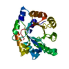
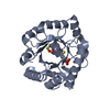
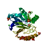
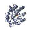
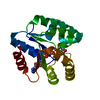
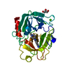
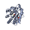
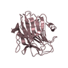
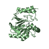
| Title | Crystal Structure of Bacillus subtilis Thiamin Phosphate Synthase Complexed with a Carboxylated Thiazole Phosphate | ||||||
 Components Components | Thiamine-phosphate pyrophosphorylase | ||||||
 Keywords Keywords |  TRANSFERASE / thiamin biosynthesis / TIM barrel / protein-intermediate complex TRANSFERASE / thiamin biosynthesis / TIM barrel / protein-intermediate complex | ||||||
| Function / homology |  Function and homology information Function and homology informationthiamine phosphate synthase / thiamine-phosphate diphosphorylase activity / thiamine diphosphate biosynthetic process / thiamine biosynthetic process / magnesium ion binding / cytoplasmSimilarity search - Function | ||||||
| Biological species |  Bacillus subtilis (bacteria) Bacillus subtilis (bacteria) | ||||||
| Method | X-RAY DIFFRACTION / SYNCHROTRON / MOLECULAR REPLACEMENT / molecular replacement / Resolution: 1.95 Å | ||||||
 Authors Authors | McCulloch, K.M. / Hanes, J.W. / Abdelwahed, S. / Mahanta, N. / Hazra, A. / Ishida, K. / Begley, T.P. / Ealick, S.E. | ||||||
 Citation Citation | Journal: to be published Title: Crystal Structure and Kinetic Characterization of Bacillus subtilis Thiamin Phosphate Synthase with a Carboxylated Thiazole Phosphate Authors: McCulloch, K.M. / Hanes, J.W. / Abdelwahed, S. / Mahanta, N. / Hazra, A. / Ishida, K. / Begley, T.P. / Ealick, S.E. | ||||||
| History |
|
- Structure visualization
| Structure viewer | Molecule: MolmilJmol/JSmol |
|---|
- Downloads & links
-Download
| PDBx/mmCIF format | 3o15.cif.gz | 60 KB | Display | PDBx/mmCIF format |
|---|---|---|---|---|
| PDB format | pdb3o15.ent.gz | 43.1 KB | Display | PDB format |
| PDBx/mmJSON format | 3o15.json.gz | Tree view | PDBx/mmJSON format | |
| Others |  Other downloads Other downloads |
-Validation report
| Arichive directory | https://data.pdbj.org/pub/pdb/validation_reports/o1/3o15ftp://data.pdbj.org/pub/pdb/validation_reports/o1/3o15 | HTTPS FTP |
|---|
-Related structure data


-Links
 PDBj
PDBj


- Assembly
| Deposited unit | 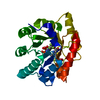
| ||||||||
|---|---|---|---|---|---|---|---|---|---|
| 1 |
| ||||||||
| Unit cell |
|
-Components
| #1: Protein | Mass: 25297.752 Da / Num. of mol.: 1 Source method: isolated from a genetically manipulated source Source: (gene. exp.) Bacillus subtilis (bacteria) / Gene: thiE / Plasmid: pQE-32 / Production host: Escherichia coli (E. coli) / Strain (production host): SG13009 / References: UniProt: P39594, thiamine phosphate synthase |
|---|---|
| #2: Chemical | ChemComp-3NM /   Mass: 267.196 Da / Num. of mol.: 1 / Source method: obtained synthetically / Formula: C7H10NO6PS Mass: 267.196 Da / Num. of mol.: 1 / Source method: obtained synthetically / Formula: C7H10NO6PS |
| #3: Chemical | ChemComp-IFP /   Mass: 175.111 Da / Num. of mol.: 1 / Source method: obtained synthetically / Formula: C6H4F3N3 Mass: 175.111 Da / Num. of mol.: 1 / Source method: obtained synthetically / Formula: C6H4F3N3 |
| #4: Chemical | ChemComp-POP / Pyrophosphate  Mass: 175.959 Da / Num. of mol.: 1 / Source method: obtained synthetically / Formula: H2O7P2 Mass: 175.959 Da / Num. of mol.: 1 / Source method: obtained synthetically / Formula: H2O7P2 |
| #5: Water | ChemComp-HOH / Water Mass: 18.015 Da / Num. of mol.: 212 / Source method: isolated from a natural source / Formula: H2O Mass: 18.015 Da / Num. of mol.: 212 / Source method: isolated from a natural source / Formula: H2O |
-Experimental details
-Experiment
| Experiment | Method: X-RAY DIFFRACTION / Number of used crystals: 1 |
|---|
- Sample preparation
| Crystal | Density Matthews: 2.7 Å3/Da / Density % sol: 54.5 % |
|---|---|
| Crystal grow | Temperature: 277 K / Method: vapor diffusion, hanging drop / pH: 7.5 Details: 22-28% PEG4000, 75 mM MgCl2, 1 mM DTT, 100 mM Tris, pH 7.5, vapor diffusion, hanging drop, temperature 277K |
-Data collection
| Diffraction | Mean temperature: 100 K | |||||||||||||||||||||||||||||||||||||||||||||||||||||||||||||||||||||||||||||
|---|---|---|---|---|---|---|---|---|---|---|---|---|---|---|---|---|---|---|---|---|---|---|---|---|---|---|---|---|---|---|---|---|---|---|---|---|---|---|---|---|---|---|---|---|---|---|---|---|---|---|---|---|---|---|---|---|---|---|---|---|---|---|---|---|---|---|---|---|---|---|---|---|---|---|---|---|---|---|
| Diffraction source | Source: SYNCHROTRON / Site: APS  / Beamline: 24-ID-C / Wavelength: 0.9792 Å / Beamline: 24-ID-C / Wavelength: 0.9792 Å | |||||||||||||||||||||||||||||||||||||||||||||||||||||||||||||||||||||||||||||
| Detector | Type: ADSC QUANTUM 315 / Detector: CCD / Date: Aug 18, 2009 | |||||||||||||||||||||||||||||||||||||||||||||||||||||||||||||||||||||||||||||
| Radiation | Protocol: SINGLE WAVELENGTH / Monochromatic (M) / Laue (L): M / Scattering type: x-ray | |||||||||||||||||||||||||||||||||||||||||||||||||||||||||||||||||||||||||||||
| Radiation wavelength | Wavelength: 0.9792 Å / Relative weight: 1 | |||||||||||||||||||||||||||||||||||||||||||||||||||||||||||||||||||||||||||||
| Reflection | Resolution: 1.95→50 Å / Num. obs: 20400 / % possible obs: 98.3 % / Redundancy: 3.4 % / Rmerge(I) obs: 0.062 / Χ2: 1.168 / Net I/σ(I): 18.2 | |||||||||||||||||||||||||||||||||||||||||||||||||||||||||||||||||||||||||||||
| Reflection shell |
|
-Phasing
| Phasing | Method: molecular replacement |
|---|
- Processing
| Software |
| ||||||||||||||||||||||||
|---|---|---|---|---|---|---|---|---|---|---|---|---|---|---|---|---|---|---|---|---|---|---|---|---|---|
| Refinement | Method to determine structure: MOLECULAR REPLACEMENT / Resolution: 1.95→28.35 Å / Occupancy max: 1 / Occupancy min: 1 / σ(F): 0
| ||||||||||||||||||||||||
| Solvent computation | Bsol: 56.263 Å2 | ||||||||||||||||||||||||
| Displacement parameters | Biso max: 75.26 Å2 / Biso mean: 28.8883 Å2 / Biso min: 11.83 Å2
| ||||||||||||||||||||||||
| Refinement step | Cycle: LAST / Resolution: 1.95→28.35 Å
| ||||||||||||||||||||||||
| Refine LS restraints |
| ||||||||||||||||||||||||
| Xplor file |
|
