 Movie
Movie Controller

[English] 日本語
 Yorodumi
- PDB-3baf: Crystal structure of shikimate kinase from Mycobacterium tubercul... -
+ Open data

- Basic information
| Entry | Database: PDB / ID: 3baf | ||||||
|---|---|---|---|---|---|---|---|
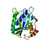
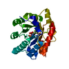
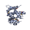
| Title | Crystal structure of shikimate kinase from Mycobacterium tuberculosis in complex with AMP-PNP | ||||||
 Components Components | Shikimate kinase | ||||||
 Keywords Keywords | TRANSFERASE / shikimate kinase / shikimate pathway / AMP-PNP / Amino-acid biosynthesis / Aromatic amino acid biosynthesis / ATP-binding / Cytoplasm / Magnesium / Metal-binding / Nucleotide-binding | ||||||
| Function / homology |  Function and homology informationshikimate kinase / shikimate metabolic process / shikimate kinase activity / Chorismate via Shikimate Pathway / chorismate biosynthetic process / aromatic amino acid family biosynthetic process / amino acid biosynthetic process / phosphorylation / magnesium ion binding / ATP binding / cytosol Function and homology informationshikimate kinase / shikimate metabolic process / shikimate kinase activity / Chorismate via Shikimate Pathway / chorismate biosynthetic process / aromatic amino acid family biosynthetic process / amino acid biosynthetic process / phosphorylation / magnesium ion binding / ATP binding / cytosolSimilarity search - Function | ||||||
| Biological species |   Mycobacterium tuberculosis (bacteria) Mycobacterium tuberculosis (bacteria) | ||||||
| Method | X-RAY DIFFRACTION / SYNCHROTRON / MOLECULAR REPLACEMENT / Resolution: 2.25 Å | ||||||
 Authors Authors | Faim, L.M. / Dias, M.V.B. / Vasconcelos, I.G. / Basso, L.A. / Santos, D.S. / Azevedo, W.F. / Ruggiero, N.J. | ||||||
 Citation Citation | Journal: To be Published Title: Crystal Structure for shikimate kinase from Mycobacterium tuberculosis in complex with AMP-PNP Authors: Faim, L.M. / Dias, M.V.B. / Vasconcelos, I.G. / Basso, L.A. / Santos, D.S. / Azevedo, W.F. / Ruggiero, N.J. | ||||||
| History |
|
- Structure visualization
| Structure viewer | Molecule: MolmilJmol/JSmol |
|---|
- Downloads & links
-Download
| PDBx/mmCIF format | 3baf.cif.gz | 49.7 KB | Display | PDBx/mmCIF format |
|---|---|---|---|---|
| PDB format | pdb3baf.ent.gz | 34.6 KB | Display | PDB format |
| PDBx/mmJSON format | 3baf.json.gz | Tree view | PDBx/mmJSON format | |
| Others |  Other downloads Other downloads |
-Validation report
| Arichive directory | https://data.pdbj.org/pub/pdb/validation_reports/ba/3bafftp://data.pdbj.org/pub/pdb/validation_reports/ba/3baf | HTTPS FTP |
|---|
-Related structure data
| Related structure data | 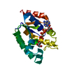 1zyuS S: Starting model for refinement |
|---|---|
| Similar structure data |


-Links
 PDBj
PDBj


- Assembly
| Deposited unit | 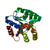
| ||||||||
|---|---|---|---|---|---|---|---|---|---|
| 1 |
| ||||||||
| Unit cell |
|
-Components
| #1: Protein | / SK Mass: 18612.352 Da / Num. of mol.: 1 Source method: isolated from a genetically manipulated source Source: (gene. exp.) Mycobacterium tuberculosis (bacteria) / Gene: aroK / Plasmid: pET23a / Production host: Escherichia coli (E. coli)References: UniProt: P0A4Z2, UniProt: P9WPY3*PLUS, shikimate kinase |
|---|---|
| #2: Chemical | ChemComp-ANP /   Mass: 506.196 Da / Num. of mol.: 1 / Source method: obtained synthetically / Formula: C10H17N6O12P3 / Comment: AMP-PNP, energy-carrying molecule analogue*YM Mass: 506.196 Da / Num. of mol.: 1 / Source method: obtained synthetically / Formula: C10H17N6O12P3 / Comment: AMP-PNP, energy-carrying molecule analogue*YM |
| #3: Chemical | ChemComp-SKM / (Shikimic acid  Mass: 174.151 Da / Num. of mol.: 1 / Source method: obtained synthetically / Formula: C7H10O5 Mass: 174.151 Da / Num. of mol.: 1 / Source method: obtained synthetically / Formula: C7H10O5 |
| #4: Water | ChemComp-HOH / Water Mass: 18.015 Da / Num. of mol.: 136 / Source method: isolated from a natural source / Formula: H2O Mass: 18.015 Da / Num. of mol.: 136 / Source method: isolated from a natural source / Formula: H2O |
-Experimental details
-Experiment
| Experiment | Method: X-RAY DIFFRACTION / Number of used crystals: 1 |
|---|
- Sample preparation
| Crystal | Density Matthews: 3.43 Å3/Da / Density % sol: 64.15 % |
|---|---|
| Crystal grow | Temperature: 293 K / Method: vapor diffusion / pH: 8 / Details: PEG, pH8, VAPOR DIFFUSION, temperature 293K |
-Data collection
| Diffraction | Mean temperature: 100 K |
|---|---|
| Diffraction source | Source: SYNCHROTRON / Site: LNLS  / Beamline: D03B-MX1 / Wavelength: 1.427 Å / Beamline: D03B-MX1 / Wavelength: 1.427 Å |
| Detector | Type: MAR CCD 165 mm / Detector: CCD / Date: Apr 17, 2007 |
| Radiation | Monochromator: GRAPHITE / Protocol: SINGLE WAVELENGTH / Monochromatic (M) / Laue (L): M / Scattering type: x-ray |
| Radiation wavelength | Wavelength: 1.427 Å / Relative weight: 1 |
| Reflection | Resolution: 2.25→37.42 Å / Num. obs: 12600 / % possible obs: 99.8 % / Observed criterion σ(F): 2 / Observed criterion σ(I): 2 / Redundancy: 3.5 % / Biso Wilson estimate: 35.1 Å2 / Rmerge(I) obs: 0.19 / Rsym value: 0.13 / Net I/σ(I): 25.8 |
| Reflection shell | Resolution: 2.25→2.37 Å / Redundancy: 3.4 % / Rmerge(I) obs: 0.19 / Num. unique all: 1816 / % possible all: 100 |
- Processing
| Software |
| ||||||||||||||||||||||||||||||||||||||||||||||||||||||||||||||||||||||||||||||||||||||||||
|---|---|---|---|---|---|---|---|---|---|---|---|---|---|---|---|---|---|---|---|---|---|---|---|---|---|---|---|---|---|---|---|---|---|---|---|---|---|---|---|---|---|---|---|---|---|---|---|---|---|---|---|---|---|---|---|---|---|---|---|---|---|---|---|---|---|---|---|---|---|---|---|---|---|---|---|---|---|---|---|---|---|---|---|---|---|---|---|---|---|---|---|
| Refinement | Method to determine structure: MOLECULAR REPLACEMENT Starting model: PDB ENTRY 1ZYU Resolution: 2.25→37.42 Å / Cor.coef. Fo:Fc: 0.939 / Cor.coef. Fo:Fc free: 0.915 / SU B: 6.165 / SU ML: 0.155 / Cross valid method: THROUGHOUT / σ(F): 0 / σ(I): 2 / ESU R: 0.222 / ESU R Free: 0.223 / Stereochemistry target values: MAXIMUM LIKELIHOOD / Details: HYDROGENS HAVE BEEN ADDED IN THE RIDING POSITIONS
| ||||||||||||||||||||||||||||||||||||||||||||||||||||||||||||||||||||||||||||||||||||||||||
| Solvent computation | Ion probe radii: 0.8 Å / Shrinkage radii: 0.8 Å / VDW probe radii: 1.2 Å / Solvent model: MASK | ||||||||||||||||||||||||||||||||||||||||||||||||||||||||||||||||||||||||||||||||||||||||||
| Displacement parameters | Biso mean: 34.889 Å2
| ||||||||||||||||||||||||||||||||||||||||||||||||||||||||||||||||||||||||||||||||||||||||||
| Refinement step | Cycle: LAST / Resolution: 2.25→37.42 Å
| ||||||||||||||||||||||||||||||||||||||||||||||||||||||||||||||||||||||||||||||||||||||||||
| Refine LS restraints |
| ||||||||||||||||||||||||||||||||||||||||||||||||||||||||||||||||||||||||||||||||||||||||||
| LS refinement shell | Resolution: 2.25→2.308 Å / Total num. of bins used: 20
|
