 Movie
Movie Controller


+ Open data

- Basic information
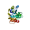
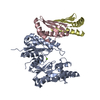
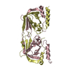
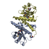
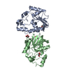
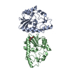
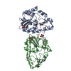
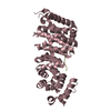
| Entry | Database: PDB / ID: 3enp | ||||||
|---|---|---|---|---|---|---|---|
| Title | Crystal structure of human cgi121 | ||||||
 Components Components | TP53RK-binding protein | ||||||
 Keywords Keywords |  HYDROLASE / KEOPS complex telomere kinase regulator / Nucleus HYDROLASE / KEOPS complex telomere kinase regulator / Nucleus | ||||||
| Function / homology |  Function and homology information Function and homology informationEKC/KEOPS complex / tRNA threonylcarbamoyladenosine modification / tRNA modification in the nucleus and cytosol / protein kinase binding / nucleus / cytosol / cytoplasmSimilarity search - Function | ||||||
| Biological species |  Homo sapiens (human) Homo sapiens (human) | ||||||
| Method | X-RAY DIFFRACTION / SYNCHROTRON / SAD / Resolution: 2.48 Å | ||||||
 Authors Authors | Haffani, Y.Z. / Ceccarelli, D.F. / Neculai, D. / Mao, D.Y. / Sicheri, F. | ||||||
 Citation Citation | Journal: Mol.Cell / Year: 2008 Title: Atomic structure of the KEOPS complex: an ancient protein kinase-containing molecular machine. Authors: Mao, D.Y. / Neculai, D. / Downey, M. / Orlicky, S. / Haffani, Y.Z. / Ceccarelli, D.F. / Ho, J.S. / Szilard, R.K. / Zhang, W. / Ho, C.S. / Wan, L. / Fares, C. / Rumpel, S. / Kurinov, I. / ...Authors: Mao, D.Y. / Neculai, D. / Downey, M. / Orlicky, S. / Haffani, Y.Z. / Ceccarelli, D.F. / Ho, J.S. / Szilard, R.K. / Zhang, W. / Ho, C.S. / Wan, L. / Fares, C. / Rumpel, S. / Kurinov, I. / Arrowsmith, C.H. / Durocher, D. / Sicheri, F. | ||||||
| History |
|
- Structure visualization
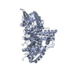
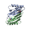
| Structure viewer | Molecule: MolmilJmol/JSmol |
|---|
- Downloads & links
-Download
| PDBx/mmCIF format | 3enp.cif.gz | 74.1 KB | Display | PDBx/mmCIF format |
|---|---|---|---|---|
| PDB format | pdb3enp.ent.gz | 60.9 KB | Display | PDB format |
| PDBx/mmJSON format | 3enp.json.gz | Tree view | PDBx/mmJSON format | |
| Others |  Other downloads Other downloads |
-Validation report
| Arichive directory | https://data.pdbj.org/pub/pdb/validation_reports/en/3enpftp://data.pdbj.org/pub/pdb/validation_reports/en/3enp | HTTPS FTP |
|---|
-Related structure data

-Links
 PDBj
PDBj- Assembly
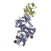
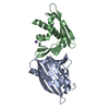
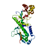
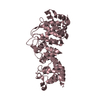
| Deposited unit | 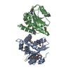
| ||||||||
|---|---|---|---|---|---|---|---|---|---|
| 1 |
| ||||||||
| 2 | 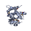
| ||||||||
| 3 | 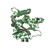
| ||||||||
| Unit cell |
|
-Components
| #1: Protein | Mass: 20046.512 Da / Num. of mol.: 2 Source method: isolated from a genetically manipulated source Source: (gene. exp.) Homo sapiens (human) / Gene: CGI-121, My019, TPRKB / Plasmid: pGEX-4T3 / Production host:  Escherichia coli (E. coli) / Strain (production host): BL21-DE3 / References: UniProt: Q9Y3C4 Escherichia coli (E. coli) / Strain (production host): BL21-DE3 / References: UniProt: Q9Y3C4#2: Water | ChemComp-HOH / | Water Mass: 18.015 Da / Num. of mol.: 18 / Source method: isolated from a natural source / Formula: H2O Mass: 18.015 Da / Num. of mol.: 18 / Source method: isolated from a natural source / Formula: H2O |
|---|
-Experimental details
-Experiment
| Experiment | Method: X-RAY DIFFRACTION / Number of used crystals: 1 |
|---|
- Sample preparation
| Crystal | Density Matthews: 2.76 Å3/Da / Density % sol: 55.51 % |
|---|---|
| Crystal grow | Temperature: 277 K / Method: vapor diffusion, hanging drop / pH: 8 Details: 20% PEG 1500, 0.2 M CaOAc, 0.1 M imidazole, pH 8, VAPOR DIFFUSION, HANGING DROP, temperature 277K |
-Data collection
| Diffraction | Mean temperature: 100 K |
|---|---|
| Diffraction source | Source: SYNCHROTRON / Site: APS  / Beamline: 14-BM-D / Wavelength: 0.9792 Å / Beamline: 14-BM-D / Wavelength: 0.9792 Å |
| Detector | Type: ADSC QUANTUM 315 / Detector: CCD / Date: Apr 10, 2004 / Details: mirrors |
| Radiation | Monochromator: SI(111) / Protocol: SINGLE WAVELENGTH / Monochromatic (M) / Laue (L): M / Scattering type: x-ray |
| Radiation wavelength | Wavelength: 0.9792 Å / Relative weight: 1 |
| Reflection | Resolution: 2.48→31.88 Å / Num. all: 13866 / Num. obs: 13866 / % possible obs: 99.8 % / Observed criterion σ(F): 0 / Observed criterion σ(I): 2 / Redundancy: 6.62 % / Rmerge(I) obs: 0.0494 / Net I/σ(I): 15.01 |
| Reflection shell | Resolution: 2.48→2.58 Å / Redundancy: 5.9 % / Rmerge(I) obs: 0.25 / Mean I/σ(I) obs: 2.53 / % possible all: 99.6 |
- Processing
| Software |
| ||||||||||||||||||||||||||||||||||||||||||||||||||||||||||||||||||||||||||||||||||||||||||||||||||||||||||||||||||||||||||||||||||||||||||||||||||||||||||||||||||||||||||
|---|---|---|---|---|---|---|---|---|---|---|---|---|---|---|---|---|---|---|---|---|---|---|---|---|---|---|---|---|---|---|---|---|---|---|---|---|---|---|---|---|---|---|---|---|---|---|---|---|---|---|---|---|---|---|---|---|---|---|---|---|---|---|---|---|---|---|---|---|---|---|---|---|---|---|---|---|---|---|---|---|---|---|---|---|---|---|---|---|---|---|---|---|---|---|---|---|---|---|---|---|---|---|---|---|---|---|---|---|---|---|---|---|---|---|---|---|---|---|---|---|---|---|---|---|---|---|---|---|---|---|---|---|---|---|---|---|---|---|---|---|---|---|---|---|---|---|---|---|---|---|---|---|---|---|---|---|---|---|---|---|---|---|---|---|---|---|---|---|---|---|---|
| Refinement | Method to determine structure: SAD / Resolution: 2.48→30.61 Å / Cor.coef. Fo:Fc: 0.949 / Cor.coef. Fo:Fc free: 0.929 / SU B: 25.581 / SU ML: 0.258 / TLS residual ADP flag: LIKELY RESIDUAL / Cross valid method: THROUGHOUT / ESU R: 0.434 / ESU R Free: 0.277 / Stereochemistry target values: MAXIMUM LIKELIHOOD / Details: HYDROGENS HAVE BEEN ADDED IN THE RIDING POSITIONS
| ||||||||||||||||||||||||||||||||||||||||||||||||||||||||||||||||||||||||||||||||||||||||||||||||||||||||||||||||||||||||||||||||||||||||||||||||||||||||||||||||||||||||||
| Solvent computation | Ion probe radii: 0.8 Å / Shrinkage radii: 0.8 Å / VDW probe radii: 1.2 Å / Solvent model: MASK | ||||||||||||||||||||||||||||||||||||||||||||||||||||||||||||||||||||||||||||||||||||||||||||||||||||||||||||||||||||||||||||||||||||||||||||||||||||||||||||||||||||||||||
| Displacement parameters | Biso mean: 54.801 Å2
| ||||||||||||||||||||||||||||||||||||||||||||||||||||||||||||||||||||||||||||||||||||||||||||||||||||||||||||||||||||||||||||||||||||||||||||||||||||||||||||||||||||||||||
| Refinement step | Cycle: LAST / Resolution: 2.48→30.61 Å
| ||||||||||||||||||||||||||||||||||||||||||||||||||||||||||||||||||||||||||||||||||||||||||||||||||||||||||||||||||||||||||||||||||||||||||||||||||||||||||||||||||||||||||
| Refine LS restraints |
| ||||||||||||||||||||||||||||||||||||||||||||||||||||||||||||||||||||||||||||||||||||||||||||||||||||||||||||||||||||||||||||||||||||||||||||||||||||||||||||||||||||||||||
| LS refinement shell | Resolution: 2.48→2.544 Å / Total num. of bins used: 20
| ||||||||||||||||||||||||||||||||||||||||||||||||||||||||||||||||||||||||||||||||||||||||||||||||||||||||||||||||||||||||||||||||||||||||||||||||||||||||||||||||||||||||||
| Refinement TLS params. | Method: refined / Refine-ID: X-RAY DIFFRACTION
| ||||||||||||||||||||||||||||||||||||||||||||||||||||||||||||||||||||||||||||||||||||||||||||||||||||||||||||||||||||||||||||||||||||||||||||||||||||||||||||||||||||||||||
| Refinement TLS group |
|
