 Movie
Movie Controller


+ Open data

- Basic information
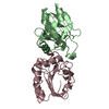
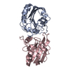
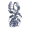
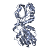
| Entry | Database: PDB / ID: 3mng | ||||||
|---|---|---|---|---|---|---|---|
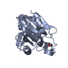
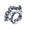
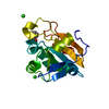
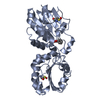
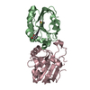
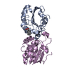
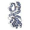
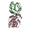
| Title | wild type human PrxV with DTT bound as a competitive inhibitor | ||||||
 Components Components | Peroxiredoxin-5, mitochondrial | ||||||
 Keywords Keywords | OXIDOREDUCTASE / peroxiredoxin / peroxidase / PrxV / substrate analog / DTT | ||||||
| Function / homology |  Function and homology information Function and homology informationperoxynitrite reductase activity / reactive nitrogen species metabolic process / negative regulation of transcription by RNA polymerase III / negative regulation of oxidoreductase activity / NADPH oxidation / regulation of apoptosis involved in tissue homeostasis / RNA polymerase III transcription regulatory region sequence-specific DNA binding / thioredoxin-dependent peroxiredoxin / thioredoxin peroxidase activity / Detoxification of Reactive Oxygen Species ...peroxynitrite reductase activity / reactive nitrogen species metabolic process / negative regulation of transcription by RNA polymerase III / negative regulation of oxidoreductase activity / NADPH oxidation / regulation of apoptosis involved in tissue homeostasis / RNA polymerase III transcription regulatory region sequence-specific DNA binding / thioredoxin-dependent peroxiredoxin / thioredoxin peroxidase activity / Detoxification of Reactive Oxygen Species / antioxidant activity / cysteine-type endopeptidase inhibitor activity involved in apoptotic process / peroxisomal matrix / positive regulation of collagen biosynthetic process / cell redox homeostasis / hydrogen peroxide catabolic process / TP53 Regulates Metabolic Genes / peroxidase activity / cellular response to reactive oxygen species / peroxisome / cellular response to oxidative stress / cytoplasmic vesicle / response to oxidative stress / mitochondrial matrix / inflammatory response / signaling receptor binding / intracellular membrane-bounded organelle / negative regulation of apoptotic process / perinuclear region of cytoplasm / mitochondrion / extracellular space / extracellular exosome / identical protein binding / nucleus / cytosol / cytoplasmSimilarity search - Function | ||||||
| Biological species |  Homo sapiens (human) Homo sapiens (human) | ||||||
| Method | X-RAY DIFFRACTION / SYNCHROTRON / Rigid body refinement of starting model / Resolution: 1.45 Å | ||||||
 Authors Authors | Hall, A. / Karplus, P.A. | ||||||
 Citation Citation | Journal: J.Mol.Biol. / Year: 2010 Title: Structural Evidence that Peroxiredoxin Catalytic Power Is Based on Transition-State Stabilization. Authors: Hall, A. / Parsonage, D. / Poole, L.B. / Karplus, P.A. | ||||||
| History |
|
- Structure visualization
| Structure viewer | Molecule: MolmilJmol/JSmol |
|---|
- Downloads & links
-Download
| PDBx/mmCIF format | 3mng.cif.gz | 90.1 KB | Display | PDBx/mmCIF format |
|---|---|---|---|---|
| PDB format | pdb3mng.ent.gz | 67.4 KB | Display | PDB format |
| PDBx/mmJSON format | 3mng.json.gz | Tree view | PDBx/mmJSON format | |
| Others |  Other downloads Other downloads |
-Validation report
| Arichive directory | https://data.pdbj.org/pub/pdb/validation_reports/mn/3mngftp://data.pdbj.org/pub/pdb/validation_reports/mn/3mng | HTTPS FTP |
|---|
-Related structure data
| Related structure data | 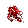 1hd2S S: Starting model for refinement |
|---|---|
| Similar structure data |


-Links
 PDBj
PDBj


- Assembly
| Deposited unit | 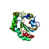
| ||||||||
|---|---|---|---|---|---|---|---|---|---|
| 1 | 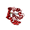
| ||||||||
| Unit cell |
| ||||||||
| Components on special symmetry positions |
|
-Components
| #1: Protein | / Peroxiredoxin V / Prx-V / Peroxisomal antioxidant enzyme / PLP / Thioredoxin reductase / ...Peroxiredoxin V / Prx-V / Peroxisomal antioxidant enzyme / PLP / Thioredoxin reductase / Thioredoxin peroxidase PMP20 / Antioxidant enzyme B166 / AOEB166 / TPx type VI / Liver tissue 2D-page spot 71B / Alu corepressor 1 Mass: 18325.045 Da / Num. of mol.: 1 Source method: isolated from a genetically manipulated source Source: (gene. exp.) Homo sapiens (human) / Gene: ACR1, aoeb166, arc1, pmp20, PRDX5, SBBI10 / Plasmid: pQE30-Prx5 / Production host:  Escherichia coli (E. coli) / Strain (production host): B834/pREP4 / References: UniProt: P30044, peroxiredoxin Escherichia coli (E. coli) / Strain (production host): B834/pREP4 / References: UniProt: P30044, peroxiredoxin | ||||
|---|---|---|---|---|---|
| #2: Chemical | ChemComp-D1D / (  Mass: 152.235 Da / Num. of mol.: 1 / Source method: obtained synthetically / Formula: C4H8O2S2 Mass: 152.235 Da / Num. of mol.: 1 / Source method: obtained synthetically / Formula: C4H8O2S2 | ||||
| #3: Chemical | ChemComp-BR / Bromide  Mass: 79.904 Da / Num. of mol.: 8 / Source method: obtained synthetically / Formula: Br Mass: 79.904 Da / Num. of mol.: 8 / Source method: obtained synthetically / Formula: Br#4: Chemical | Glycerol  Mass: 92.094 Da / Num. of mol.: 2 / Source method: obtained synthetically / Formula: C3H8O3 Mass: 92.094 Da / Num. of mol.: 2 / Source method: obtained synthetically / Formula: C3H8O3#5: Water | ChemComp-HOH / | Water Mass: 18.015 Da / Num. of mol.: 288 / Source method: isolated from a natural source / Formula: H2O Mass: 18.015 Da / Num. of mol.: 288 / Source method: isolated from a natural source / Formula: H2O |
-Experimental details
-Experiment
| Experiment | Method: X-RAY DIFFRACTION / Number of used crystals: 2 |
|---|
- Sample preparation
| Crystal | Density Matthews: 3.83 Å3/Da / Density % sol: 67.87 % |
|---|---|
| Crystal grow | Temperature: 298 K / Method: vapor diffusion, hanging drop / pH: 5.6 Details: Reservoir contained 1.6 M ammonium sulfate, 0.1 M sodium citrate, 0.2 M sodium/potassium tartrate. Protein was stored at 0.8 mM with 10 mM DTT., pH 5.6, VAPOR DIFFUSION, HANGING DROP, temperature 298K |
-Data collection
| Diffraction | Mean temperature: 100 K |
|---|---|
| Diffraction source | Source: SYNCHROTRON / Site: ALS  / Beamline: 5.0.1 / Wavelength: 0.978 Å / Beamline: 5.0.1 / Wavelength: 0.978 Å |
| Detector | Type: ADSC QUANTUM 210 / Detector: CCD / Date: Dec 18, 2009 |
| Radiation | Monochromator: Single crystal, cylindrically bent Si(220) / Protocol: SINGLE WAVELENGTH / Monochromatic (M) / Laue (L): M / Scattering type: x-ray |
| Radiation wavelength | Wavelength: 0.978 Å / Relative weight: 1 |
| Reflection | Resolution: 1.45→59 Å / Num. obs: 48609 / % possible obs: 99.98 % / Redundancy: 36.7 % / Rsym value: 0.094 / Net I/σ(I): 30.9 |
| Reflection shell | Resolution: 1.45→1.53 Å / Redundancy: 14 % / Mean I/σ(I) obs: 6.9 / Num. unique all: 7365 / Rsym value: 0.393 / % possible all: 100 |
- Processing
| Software |
| ||||||||||||||||||||||||||||||||||||||||||||||||||||||||||||||||||||||||||||||||||||||||||
|---|---|---|---|---|---|---|---|---|---|---|---|---|---|---|---|---|---|---|---|---|---|---|---|---|---|---|---|---|---|---|---|---|---|---|---|---|---|---|---|---|---|---|---|---|---|---|---|---|---|---|---|---|---|---|---|---|---|---|---|---|---|---|---|---|---|---|---|---|---|---|---|---|---|---|---|---|---|---|---|---|---|---|---|---|---|---|---|---|---|---|---|
| Refinement | Method to determine structure: Rigid body refinement of starting model Starting model: pdb code 1HD2 with benzoate removed Resolution: 1.45→59 Å / Cor.coef. Fo:Fc: 0.978 / Cor.coef. Fo:Fc free: 0.969 / SU B: 0.923 / SU ML: 0.016 / Cross valid method: THROUGHOUT / ESU R: 0.035 / ESU R Free: 0.036 / Stereochemistry target values: MAXIMUM LIKELIHOOD / Details: HYDROGENS HAVE BEEN ADDED IN THE RIDING POSITIONS
| ||||||||||||||||||||||||||||||||||||||||||||||||||||||||||||||||||||||||||||||||||||||||||
| Solvent computation | Ion probe radii: 0.8 Å / Shrinkage radii: 0.8 Å / VDW probe radii: 1.4 Å / Solvent model: MASK | ||||||||||||||||||||||||||||||||||||||||||||||||||||||||||||||||||||||||||||||||||||||||||
| Displacement parameters | Biso mean: 11.049 Å2
| ||||||||||||||||||||||||||||||||||||||||||||||||||||||||||||||||||||||||||||||||||||||||||
| Refinement step | Cycle: LAST / Resolution: 1.45→59 Å
| ||||||||||||||||||||||||||||||||||||||||||||||||||||||||||||||||||||||||||||||||||||||||||
| Refine LS restraints |
| ||||||||||||||||||||||||||||||||||||||||||||||||||||||||||||||||||||||||||||||||||||||||||
| LS refinement shell | Resolution: 1.45→1.488 Å / Total num. of bins used: 20
|
