 Movie
Movie Controller


+ Open data

- Basic information
| Entry | Database: PDB / ID: 2wfc | ||||||
|---|---|---|---|---|---|---|---|
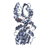
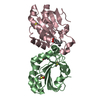
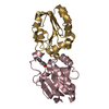
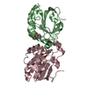
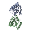
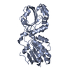
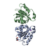
| Title | Crystal structure of peroxiredoxin 5 from Arenicola Marina | ||||||
 Components Components | PEROXIREDOXIN 5 | ||||||
 Keywords Keywords | OXIDOREDUCTASE / ANTIOXIDANT ENZYMES | ||||||
| Function / homology |  Function and homology information Function and homology informationthioredoxin-dependent peroxiredoxin / peroxisomal matrix / cell redox homeostasis / peroxidase activitySimilarity search - Function | ||||||
| Biological species |  ARENICOLA MARINA (lugworm) ARENICOLA MARINA (lugworm) | ||||||
| Method | X-RAY DIFFRACTION / SYNCHROTRON / MOLECULAR REPLACEMENT / Resolution: 1.75 Å | ||||||
 Authors Authors | Smeets, A. / Declercq, J.P. | ||||||
 Citation Citation | Journal: To be Published Title: Crystal Structure of Peroxiredoxin 5 from Arenicola Marina Authors: Smeets, A. / Knoops, B. / Declercq, J.P. | ||||||
| History |
| ||||||
| Remark 650 | HELIX DETERMINATION METHOD: AUTHOR PROVIDED |
- Structure visualization
| Structure viewer | Molecule: MolmilJmol/JSmol |
|---|
- Downloads & links
-Download
| PDBx/mmCIF format | 2wfc.cif.gz | 140.1 KB | Display | PDBx/mmCIF format |
|---|---|---|---|---|
| PDB format | pdb2wfc.ent.gz | 110.5 KB | Display | PDB format |
| PDBx/mmJSON format | 2wfc.json.gz | Tree view | PDBx/mmJSON format | |
| Others |  Other downloads Other downloads |
-Validation report
| Arichive directory | https://data.pdbj.org/pub/pdb/validation_reports/wf/2wfcftp://data.pdbj.org/pub/pdb/validation_reports/wf/2wfc | HTTPS FTP |
|---|
-Related structure data
| Related structure data | 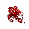 1hd2S S: Starting model for refinement |
|---|---|
| Similar structure data |


-Links
 PDBj
PDBj- Assembly
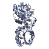
| Deposited unit | 
| ||||||||||||||||
|---|---|---|---|---|---|---|---|---|---|---|---|---|---|---|---|---|---|
| 1 | 
| ||||||||||||||||
| 2 | 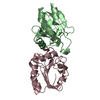
| ||||||||||||||||
| Unit cell |
| ||||||||||||||||
| Noncrystallographic symmetry (NCS) | NCS oper:
|
-Components
| #1: Protein | / PRDX5 Mass: 17516.062 Da / Num. of mol.: 4 / Fragment: RESIDUES 28-186 / Mutation: YES Source method: isolated from a genetically manipulated source Source: (gene. exp.) ARENICOLA MARINA (lugworm) / Plasmid: PQE-60 / Production host:  ESCHERICHIA COLI (E. coli) / Strain (production host): M15 / References: UniProt: Q1AN23, peroxiredoxin ESCHERICHIA COLI (E. coli) / Strain (production host): M15 / References: UniProt: Q1AN23, peroxiredoxin#2: Chemical | ChemComp-ACT / | Acetate  Mass: 59.044 Da / Num. of mol.: 1 / Source method: obtained synthetically / Formula: C2H3O2 Mass: 59.044 Da / Num. of mol.: 1 / Source method: obtained synthetically / Formula: C2H3O2#3: Water | ChemComp-HOH / | Water Mass: 18.015 Da / Num. of mol.: 627 / Source method: isolated from a natural source / Formula: H2O Mass: 18.015 Da / Num. of mol.: 627 / Source method: isolated from a natural source / Formula: H2OCompound details | ENGINEERED RESIDUE IN CHAIN A, CYS 74 TO SER ENGINEERED RESIDUE IN CHAIN B, CYS 74 TO SER ...ENGINEERED | |
|---|
-Experimental details
-Experiment
| Experiment | Method: X-RAY DIFFRACTION / Number of used crystals: 1 |
|---|
- Sample preparation
| Crystal | Density Matthews: 1.98 Å3/Da / Density % sol: 38 % / Description: NONE |
|---|---|
| Crystal grow | Details: PEG 4000 29%(W/V), DTT 0.001 M |
-Data collection
| Diffraction | Mean temperature: 100 K |
|---|---|
| Diffraction source | Source: SYNCHROTRON / Site: ESRF  / Beamline: BM30A / Wavelength: 0.9797 / Beamline: BM30A / Wavelength: 0.9797 |
| Detector | Type: ADSC CCD / Detector: CCD / Details: SECOND CRYSTAL SAGITALLY BENT |
| Radiation | Monochromator: FIRST CRYSTAL FLAT AND N2 COOLED / Protocol: SINGLE WAVELENGTH / Monochromatic (M) / Laue (L): M / Scattering type: x-ray |
| Radiation wavelength | Wavelength: 0.9797 Å / Relative weight: 1 |
| Reflection | Resolution: 1.75→34.17 Å / Num. obs: 51140 / % possible obs: 95.6 % / Observed criterion σ(I): 0 / Redundancy: 3.7 % / Biso Wilson estimate: 24.2 Å2 / Rmerge(I) obs: 0.05 / Net I/σ(I): 17.7 |
| Reflection shell | Resolution: 1.75→1.79 Å / Redundancy: 3.4 % / Rmerge(I) obs: 0.27 / Mean I/σ(I) obs: 4.4 / % possible all: 82.5 |
- Processing
| Software |
| ||||||||||||||||||||||||||||||||||||||||||||||||||||||||||||||||||||||||||||||||||||||||||||||||||||||||||||||||||||||||||||||||||||||||||||||||||||||||||||||||||||||||||||||||||||||
|---|---|---|---|---|---|---|---|---|---|---|---|---|---|---|---|---|---|---|---|---|---|---|---|---|---|---|---|---|---|---|---|---|---|---|---|---|---|---|---|---|---|---|---|---|---|---|---|---|---|---|---|---|---|---|---|---|---|---|---|---|---|---|---|---|---|---|---|---|---|---|---|---|---|---|---|---|---|---|---|---|---|---|---|---|---|---|---|---|---|---|---|---|---|---|---|---|---|---|---|---|---|---|---|---|---|---|---|---|---|---|---|---|---|---|---|---|---|---|---|---|---|---|---|---|---|---|---|---|---|---|---|---|---|---|---|---|---|---|---|---|---|---|---|---|---|---|---|---|---|---|---|---|---|---|---|---|---|---|---|---|---|---|---|---|---|---|---|---|---|---|---|---|---|---|---|---|---|---|---|---|---|---|---|
| Refinement | Method to determine structure: MOLECULAR REPLACEMENT Starting model: PDB ENTRY 1HD2 Resolution: 1.75→68.36 Å / Cor.coef. Fo:Fc: 0.96 / Cor.coef. Fo:Fc free: 0.937 / SU B: 5.654 / SU ML: 0.082 / Cross valid method: THROUGHOUT / ESU R: 0.128 / ESU R Free: 0.127 / Stereochemistry target values: MAXIMUM LIKELIHOOD / Details: HYDROGENS HAVE BEEN ADDED IN THE RIDING POSITIONS.
| ||||||||||||||||||||||||||||||||||||||||||||||||||||||||||||||||||||||||||||||||||||||||||||||||||||||||||||||||||||||||||||||||||||||||||||||||||||||||||||||||||||||||||||||||||||||
| Solvent computation | Ion probe radii: 0.8 Å / Shrinkage radii: 0.8 Å / VDW probe radii: 1.4 Å / Solvent model: MASK | ||||||||||||||||||||||||||||||||||||||||||||||||||||||||||||||||||||||||||||||||||||||||||||||||||||||||||||||||||||||||||||||||||||||||||||||||||||||||||||||||||||||||||||||||||||||
| Displacement parameters | Biso mean: 20.57 Å2
| ||||||||||||||||||||||||||||||||||||||||||||||||||||||||||||||||||||||||||||||||||||||||||||||||||||||||||||||||||||||||||||||||||||||||||||||||||||||||||||||||||||||||||||||||||||||
| Refinement step | Cycle: LAST / Resolution: 1.75→68.36 Å
| ||||||||||||||||||||||||||||||||||||||||||||||||||||||||||||||||||||||||||||||||||||||||||||||||||||||||||||||||||||||||||||||||||||||||||||||||||||||||||||||||||||||||||||||||||||||
| Refine LS restraints |
|