 Movie
Movie Controller

[English] 日本語
 Yorodumi
- PDB-5k2j: Crystal structure of reduced Prx3 in complex with h2o2 from Vibri... -
+ Open data

- Basic information
| Entry | Database: PDB / ID: 5k2j | ||||||
|---|---|---|---|---|---|---|---|
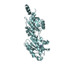
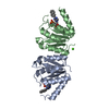
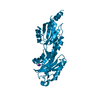
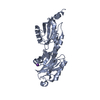
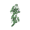
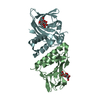
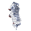
| Title | Crystal structure of reduced Prx3 in complex with h2o2 from Vibrio vulnificus | ||||||
 Components Components | 1-Cys peroxiredoxin | ||||||
 Keywords Keywords |  OXIDOREDUCTASE / 1-Cys Prx / Vibrio vulnificus / hydrogen peroxide / nitric oxide OXIDOREDUCTASE / 1-Cys Prx / Vibrio vulnificus / hydrogen peroxide / nitric oxide | ||||||
| Function / homology |  Function and homology information Function and homology informationglutathione-dependent peroxiredoxin / thioredoxin peroxidase activity / cellular response to oxidative stress Similarity search - Function | ||||||
| Biological species |  Vibrio vulnificus MO6-24/O (bacteria) Vibrio vulnificus MO6-24/O (bacteria) | ||||||
| Method | X-RAY DIFFRACTION / SYNCHROTRON / MOLECULAR REPLACEMENT / Resolution: 1.908 Å | ||||||
 Authors Authors | Ahn, J. / Ha, N.-C. | ||||||
 Citation Citation | Journal: IUCrJ / Year: 2018 Title: Crystal structure of peroxiredoxin 3 fromVibrio vulnificusand its implications for scavenging peroxides and nitric oxide. Authors: Ahn, J. / Jang, K.K. / Jo, I. / Nurhasni, H. / Lim, J.G. / Yoo, J.W. / Choi, S.H. / Ha, N.C. | ||||||
| History |
|
- Structure visualization
| Structure viewer | Molecule: MolmilJmol/JSmol |
|---|
- Downloads & links
-Download
| PDBx/mmCIF format | 5k2j.cif.gz | 369.9 KB | Display | PDBx/mmCIF format |
|---|---|---|---|---|
| PDB format | pdb5k2j.ent.gz | 302.2 KB | Display | PDB format |
| PDBx/mmJSON format | 5k2j.json.gz | Tree view | PDBx/mmJSON format | |
| Others |  Other downloads Other downloads |
-Validation report
| Arichive directory | https://data.pdbj.org/pub/pdb/validation_reports/k2/5k2jftp://data.pdbj.org/pub/pdb/validation_reports/k2/5k2j | HTTPS FTP |
|---|
-Related structure data
| Related structure data |  5k1gSC 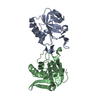 5k2iC S: Starting model for refinement C: citing same article ( |
|---|---|
| Similar structure data |

-Links
 PDBj
PDBj- Assembly
| Deposited unit | 
| ||||||||
|---|---|---|---|---|---|---|---|---|---|
| 1 | 
| ||||||||
| 2 | 
| ||||||||
| 3 | 
| ||||||||
| 4 | 
| ||||||||
| 5 | 
| ||||||||
| 6 | 
| ||||||||
| Unit cell |
|
-Components
| #1: Protein | Mass: 17545.971 Da / Num. of mol.: 12 / Mutation: C48D, C73S Source method: isolated from a genetically manipulated source Source: (gene. exp.) Vibrio vulnificus MO6-24/O (bacteria) / Strain: MO6-24/O / Production host: Escherichia coli (E. coli) / References: UniProt: A0A1Z0YU25*PLUS#2: Chemical | ChemComp-PEO / Hydrogen peroxide  Mass: 34.015 Da / Num. of mol.: 4 / Source method: obtained synthetically / Formula: H2O2 Mass: 34.015 Da / Num. of mol.: 4 / Source method: obtained synthetically / Formula: H2O2#3: Water | ChemComp-HOH / | Water Mass: 18.015 Da / Num. of mol.: 805 / Source method: isolated from a natural source / Formula: H2O Mass: 18.015 Da / Num. of mol.: 805 / Source method: isolated from a natural source / Formula: H2OSequence details | The reference database is GenBank ADV89163. This protein is C48D, C73S mutant, and residues 158-164 ...The reference database is GenBank ADV89163. This protein is C48D, C73S mutant, and residues 158-164 are expression tag. | |
|---|
-Experimental details
-Experiment
| Experiment | Method: X-RAY DIFFRACTION / Number of used crystals: 1 |
|---|
- Sample preparation
| Crystal | Density Matthews: 2.89 Å3/Da / Density % sol: 57.42 % |
|---|---|
| Crystal grow | Temperature: 287.15 K / Method: vapor diffusion, hanging drop / pH: 6.5 Details: 0.8 M sodium citrate, 0.1 M Tris-HCl, 0.2 M sodium chloride, 500 uM H2O2 PH range: 6.0-6.8 |
-Data collection
| Diffraction | Mean temperature: 100.15 K |
|---|---|
| Diffraction source | Source: SYNCHROTRON / Site: PAL/PLS  / Beamline: 5C (4A) / Wavelength: 0.9796 Å / Beamline: 5C (4A) / Wavelength: 0.9796 Å |
| Detector | Type: ADSC QUANTUM 315r / Detector: CCD / Date: Oct 20, 2015 |
| Radiation | Monochromator: Double mirrors / Protocol: SINGLE WAVELENGTH / Monochromatic (M) / Laue (L): M / Scattering type: x-ray |
| Radiation wavelength | Wavelength: 0.9796 Å / Relative weight: 1 |
| Reflection | Resolution: 1.9→50 Å / Num. obs: 182028 / % possible obs: 90 % / Redundancy: 3.4 % / Rmerge(I) obs: 0.06 / Net I/σ(I): 14.5 |
| Reflection shell | Resolution: 1.9→1.93 Å |
- Processing
| Software |
| |||||||||||||||||||||||||||||||||||||||||||||||||||||||||||||||||||||||||||||||||||||||||||||||||||||||||||||||||||||||||||||||||||||||||||||||||||||||||||||||||||||||||||||||||||||||||||||||||||||||||||||||||||||||||
|---|---|---|---|---|---|---|---|---|---|---|---|---|---|---|---|---|---|---|---|---|---|---|---|---|---|---|---|---|---|---|---|---|---|---|---|---|---|---|---|---|---|---|---|---|---|---|---|---|---|---|---|---|---|---|---|---|---|---|---|---|---|---|---|---|---|---|---|---|---|---|---|---|---|---|---|---|---|---|---|---|---|---|---|---|---|---|---|---|---|---|---|---|---|---|---|---|---|---|---|---|---|---|---|---|---|---|---|---|---|---|---|---|---|---|---|---|---|---|---|---|---|---|---|---|---|---|---|---|---|---|---|---|---|---|---|---|---|---|---|---|---|---|---|---|---|---|---|---|---|---|---|---|---|---|---|---|---|---|---|---|---|---|---|---|---|---|---|---|---|---|---|---|---|---|---|---|---|---|---|---|---|---|---|---|---|---|---|---|---|---|---|---|---|---|---|---|---|---|---|---|---|---|---|---|---|---|---|---|---|---|---|---|---|---|---|---|---|---|
| Refinement | Method to determine structure: MOLECULAR REPLACEMENT Starting model: 5K1G Resolution: 1.908→19.68 Å / SU ML: 0.24 / Cross valid method: FREE R-VALUE / σ(F): 2.22 / Phase error: 25.95 / Stereochemistry target values: ML
| |||||||||||||||||||||||||||||||||||||||||||||||||||||||||||||||||||||||||||||||||||||||||||||||||||||||||||||||||||||||||||||||||||||||||||||||||||||||||||||||||||||||||||||||||||||||||||||||||||||||||||||||||||||||||
| Solvent computation | Shrinkage radii: 0.9 Å / VDW probe radii: 1.11 Å / Solvent model: FLAT BULK SOLVENT MODEL | |||||||||||||||||||||||||||||||||||||||||||||||||||||||||||||||||||||||||||||||||||||||||||||||||||||||||||||||||||||||||||||||||||||||||||||||||||||||||||||||||||||||||||||||||||||||||||||||||||||||||||||||||||||||||
| Refinement step | Cycle: LAST / Resolution: 1.908→19.68 Å
| |||||||||||||||||||||||||||||||||||||||||||||||||||||||||||||||||||||||||||||||||||||||||||||||||||||||||||||||||||||||||||||||||||||||||||||||||||||||||||||||||||||||||||||||||||||||||||||||||||||||||||||||||||||||||
| Refine LS restraints |
| |||||||||||||||||||||||||||||||||||||||||||||||||||||||||||||||||||||||||||||||||||||||||||||||||||||||||||||||||||||||||||||||||||||||||||||||||||||||||||||||||||||||||||||||||||||||||||||||||||||||||||||||||||||||||
| LS refinement shell |
|
