 Movie
Movie Controller


+ Open data

- Basic information
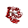
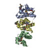
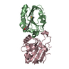
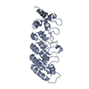
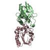
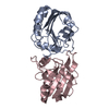
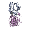
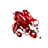
| Entry | Database: PDB / ID: 4p78 | ||||||
|---|---|---|---|---|---|---|---|
| Title | HicA3 and HicB3 toxin-antitoxin complex | ||||||
 Components Components |
| ||||||
 Keywords Keywords |  TOXIN / Yersinia pestis HicA3-HicB3 system / toxin-antitoxin TOXIN / Yersinia pestis HicA3-HicB3 system / toxin-antitoxin | ||||||
| Function / homology |  Function and homology informationendonuclease activity / sequence-specific DNA binding / mRNA binding / regulation of DNA-templated transcription Function and homology informationendonuclease activity / sequence-specific DNA binding / mRNA binding / regulation of DNA-templated transcriptionSimilarity search - Function | ||||||
| Biological species |   Yersinia pestis (bacteria) Yersinia pestis (bacteria) | ||||||
| Method | X-RAY DIFFRACTION / SYNCHROTRON / MOLECULAR REPLACEMENT / Resolution: 2.12 Å | ||||||
 Authors Authors | Li de la Sierra-Gallay, I. / Bibi-Triki, S. / van Tilbeurgh, H. / Lazar, N. / Pradel, E. | ||||||
 Citation Citation | Journal: J.Bacteriol. / Year: 2014 Title: Functional and Structural Analysis of HicA3-HicB3, a Novel Toxin-Antitoxin System of Yersinia pestis. Authors: Bibi-Triki, S. / Li de la Sierra-Gallay, I. / Lazar, N. / Leroy, A. / Van Tilbeurgh, H. / Sebbane, F. / Pradel, E. | ||||||
| History |
|
- Structure visualization
| Structure viewer | Molecule: MolmilJmol/JSmol |
|---|
- Downloads & links
-Download
| PDBx/mmCIF format | 4p78.cif.gz | 76.7 KB | Display | PDBx/mmCIF format |
|---|---|---|---|---|
| PDB format | pdb4p78.ent.gz | 55.4 KB | Display | PDB format |
| PDBx/mmJSON format | 4p78.json.gz | Tree view | PDBx/mmJSON format | |
| Others |  Other downloads Other downloads |
-Validation report
| Arichive directory | https://data.pdbj.org/pub/pdb/validation_reports/p7/4p78ftp://data.pdbj.org/pub/pdb/validation_reports/p7/4p78 | HTTPS FTP |
|---|
-Related structure data
| Related structure data |  4p7dSC  1whzS S: Starting model for refinement C: citing same article ( |
|---|---|
| Similar structure data |


-Links
 PDBj
PDBj- Assembly
| Deposited unit | 
| ||||||||
|---|---|---|---|---|---|---|---|---|---|
| 1 |
| ||||||||
| Unit cell |
|
-Components
| #1: Protein | Mass: 16334.523 Da / Num. of mol.: 2 Source method: isolated from a genetically manipulated source Source: (gene. exp.) Yersinia pestis (bacteria) / Gene: YPO3369 / Production host: Escherichia coli (E. coli) / References: UniProt: Q0WBS6, UniProt: A0A5P8YCM0*PLUS#2: Protein | Mass: 7314.652 Da / Num. of mol.: 2 Source method: isolated from a genetically manipulated source Source: (gene. exp.) Yersinia pestis (bacteria) / Gene: YP_0318 / Production host: Escherichia coli (E. coli) / References: UniProt: Q74XS2, UniProt: A0A6B3SYE0*PLUS#3: Chemical | ChemComp-GOL / | Glycerol  Mass: 92.094 Da / Num. of mol.: 1 / Source method: obtained synthetically / Formula: C3H8O3 Mass: 92.094 Da / Num. of mol.: 1 / Source method: obtained synthetically / Formula: C3H8O3#4: Water | ChemComp-HOH / | Water Mass: 18.015 Da / Num. of mol.: 115 / Source method: isolated from a natural source / Formula: H2O Mass: 18.015 Da / Num. of mol.: 115 / Source method: isolated from a natural source / Formula: H2O |
|---|
-Experimental details
-Experiment
| Experiment | Method: X-RAY DIFFRACTION |
|---|
- Sample preparation
| Crystal | Density Matthews: 1.83 Å3/Da / Density % sol: 32.95 % |
|---|---|
| Crystal grow | Temperature: 293 K / Method: vapor diffusion, sitting drop / Details: Crystals were obtained in 2.4 M di-sodium malonate |
-Data collection
| Diffraction | Mean temperature: 100 K |
|---|---|
| Diffraction source | Source: SYNCHROTRON / Site: SOLEIL  / Beamline: PROXIMA 1 / Wavelength: 0.9801 Å / Beamline: PROXIMA 1 / Wavelength: 0.9801 Å |
| Detector | Type: PSI PILATUS 6M / Detector: PIXEL / Date: Apr 10, 2013 |
| Radiation | Protocol: SINGLE WAVELENGTH / Monochromatic (M) / Laue (L): M / Scattering type: x-ray |
| Radiation wavelength | Wavelength: 0.9801 Å / Relative weight: 1 |
| Reflection | Resolution: 2.12→41.58 Å / Num. obs: 20448 / % possible obs: 99.7 % / Observed criterion σ(I): -3 / Redundancy: 5.82 % / Rmerge(I) obs: 0.11 / Net I/σ(I): 14.24 |
| Reflection shell | Resolution: 2.12→2.24 Å / Redundancy: 5.75 % / Rmerge(I) obs: 0.68 / Mean I/σ(I) obs: 2.85 / % possible all: 98.7 |
- Processing
| Software |
| ||||||||||||||||||||||||||||||||||||||||||||||||||||||||
|---|---|---|---|---|---|---|---|---|---|---|---|---|---|---|---|---|---|---|---|---|---|---|---|---|---|---|---|---|---|---|---|---|---|---|---|---|---|---|---|---|---|---|---|---|---|---|---|---|---|---|---|---|---|---|---|---|---|
| Refinement | Method to determine structure: MOLECULAR REPLACEMENT Starting model: 4P7D and 1whz Resolution: 2.12→41.58 Å / SU ML: 0.23 / Cross valid method: FREE R-VALUE / σ(F): 2 / Phase error: 21.72 / Stereochemistry target values: ML
| ||||||||||||||||||||||||||||||||||||||||||||||||||||||||
| Solvent computation | Shrinkage radii: 0.9 Å / VDW probe radii: 1.11 Å / Solvent model: FLAT BULK SOLVENT MODEL | ||||||||||||||||||||||||||||||||||||||||||||||||||||||||
| Refinement step | Cycle: LAST / Resolution: 2.12→41.58 Å
| ||||||||||||||||||||||||||||||||||||||||||||||||||||||||
| Refine LS restraints |
| ||||||||||||||||||||||||||||||||||||||||||||||||||||||||
| LS refinement shell |
|
