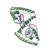
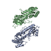
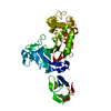
Vacuolar protein sorting-associated protein 4Vacuole
Vacuolar protein sorting-associated protein VTA1Vacuole
Keywords
PROTEIN TRANSPORT / vps4 / vta1 / AAA / ATPase / ESCRT / MVB / sorting
Function / homology
Function and homology information
ESCRT IV complex / late endosome to lysosome transport via multivesicular body sorting pathway / intralumenal vesicle formation / Endosomal Sorting Complex Required For Transport (ESCRT) / Sealing of the nuclear envelope (NE) by ESCRT-III / protein retention in Golgi apparatus / late endosome to vacuole transport via multivesicular body sorting pathway / sterol metabolic process / nuclear membrane reassembly / midbody abscission ...ESCRT IV complex / late endosome to lysosome transport via multivesicular body sorting pathway / intralumenal vesicle formation / Endosomal Sorting Complex Required For Transport (ESCRT) / Sealing of the nuclear envelope (NE) by ESCRT-III / protein retention in Golgi apparatus / late endosome to vacuole transport via multivesicular body sorting pathway / sterol metabolic process / nuclear membrane reassembly / midbody abscission / vacuole organization / multivesicular body sorting pathway / membrane fission / plasma membrane repair / late endosome to vacuole transport / multivesicular body assembly / reticulophagy / lipid transport / nucleus organization / endosomal transport / ATPase complex / ATPase activator activity / autophagosome maturation / nuclear pore / multivesicular body / macroautophagy / autophagy / protein-macromolecule adaptor activity / protein transport / midbody / endosome / endoplasmic reticulum / protein homodimerization activity / ATP hydrolysis activity / ATP binding / membrane / identical protein binding / plasma membrane / cytoplasm Similarity search - Function
Vta1/Callose synthase, N-terminal domain superfamily / Vta1/callose synthase, N-terminal / Vta1, C-terminal / Vacuolar protein sorting-associated protein Vta1-like / Vta1 like / Vta1 C-terminal domain / Vacuolar protein sorting-associated protein 4, MIT domain / MIT (microtubule interacting and transport) domain / MIT domain superfamily / Vps4 oligomerisation, C-terminal ...Vta1/Callose synthase, N-terminal domain superfamily / Vta1/callose synthase, N-terminal / Vta1, C-terminal / Vacuolar protein sorting-associated protein Vta1-like / Vta1 like / Vta1 C-terminal domain / Vacuolar protein sorting-associated protein 4, MIT domain / MIT (microtubule interacting and transport) domain / MIT domain superfamily / Vps4 oligomerisation, C-terminal / MIT domain / Microtubule Interacting and Trafficking molecule domain / Immunoglobulin FC, subunit C / Vps4 C terminal oligomerisation domain / Helicase, Ruva Protein; domain 3 - #60 / AAA ATPase, AAA+ lid domain / AAA+ lid domain / ATPase, AAA-type, conserved site / AAA-protein family signature. / Helicase, Ruva Protein; domain 3 / Single alpha-helices involved in coiled-coils or other helix-helix interfaces / ATPase family associated with various cellular activities (AAA) / ATPase, AAA-type, core / ATPases associated with a variety of cellular activities / AAA+ ATPase domain / Up-down Bundle / P-loop containing nucleoside triphosphate hydrolase / Orthogonal Bundle / Mainly Alpha Similarity search - Domain/homology
Vacuolar protein sorting-associated protein 4 / Vacuolar protein sorting-associated protein VTA1 Similarity search - Component
Resolution: 3.1→37.98 Å / Rfactor Rfree error: 0.025 / Data cutoff high absF: 1127778.72 / Data cutoff low absF: 0 / Isotropic thermal model: RESTRAINED / Cross valid method: THROUGHOUT / σ(F): 0 / Details: THe structure was refined also with refmac 5.
In the structure databanks used in Yorodumi, some data are registered as the other names, "COVID-19 virus" and "2019-nCoV". Here are the details of the virus and the list of structure data.
Jan 31, 2019. EMDB accession codes are about to change! (news from PDBe EMDB page)
EMDB accession codes are about to change! (news from PDBe EMDB page)
The allocation of 4 digits for EMDB accession codes will soon come to an end. Whilst these codes will remain in use, new EMDB accession codes will include an additional digit and will expand incrementally as the available range of codes is exhausted. The current 4-digit format prefixed with “EMD-” (i.e. EMD-XXXX) will advance to a 5-digit format (i.e. EMD-XXXXX), and so on. It is currently estimated that the 4-digit codes will be depleted around Spring 2019, at which point the 5-digit format will come into force.
The EM Navigator/Yorodumi systems omit the EMD- prefix.
Related info.:Q: What is EMD? / ID/Accession-code notation in Yorodumi/EM Navigator
Yorodumi is a browser for structure data from EMDB, PDB, SASBDB, etc.
This page is also the successor to EM Navigator detail page, and also detail information page/front-end page for Omokage search.
The word "yorodu" (or yorozu) is an old Japanese word meaning "ten thousand". "mi" (miru) is to see.
Related info.:EMDB / PDB / SASBDB / Comparison of 3 databanks / Yorodumi Search / Aug 31, 2016. New EM Navigator & Yorodumi / Yorodumi Papers / Jmol/JSmol / Function and homology information / Changes in new EM Navigator and Yorodumi
 Movie
Movie Controller


 Open data
 Basic information
 Components
 Keywords
 PROTEIN TRANSPORT / vps4 /
  Function and homology information
 Authors
 Citation
 Structure visualization
 Downloads & links
 Other downloads
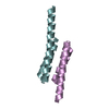


 PDBj
PDBj


 Assembly


 Sample preparation
 / Beamline: 23-ID-B / Wavelength: 0.9719 Å
 Processing