| Entry | Database: PDB / ID: 4ryk
|
|---|
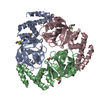
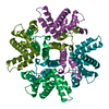
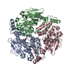
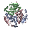
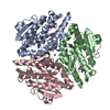
| Title | Crystal structure of a putative transcriptional regulator from Listeria monocytogenes EGD-e |
|---|
 Components Components | Lmo0325 protein |
|---|
 Keywords Keywords |  DNA BINDING PROTEIN / Structural Genomics / PSI-Biology / Midwest Center for Structural Genomics / MCSG DNA BINDING PROTEIN / Structural Genomics / PSI-Biology / Midwest Center for Structural Genomics / MCSG |
|---|
| Function / homology |  Function and homology information Function and homology information
Transcription activator MutR, C-terminal / Helix-turn-helix / Helix-turn-helix XRE-family like proteins / Cro/C1-type HTH domain profile. / Cro/C1-type helix-turn-helix domain / Lambda repressor-like, DNA-binding domain superfamily / Tetratricopeptide-like helical domain superfamilySimilarity search - Domain/homology |
|---|
| Biological species |  Listeria monocytogenes EGD-e (bacteria) Listeria monocytogenes EGD-e (bacteria) |
|---|
| Method | X-RAY DIFFRACTION / SYNCHROTRON / SAD / Resolution: 2.09 Å |
|---|
 Authors Authors | Filippova, E.V. / Wawrzak, Z. / Minasov, G. / Kiryukhina, O. / Jedrzejczak, R. / Joachimiak, A. / Anderson, W.F. / Midwest Center for Structural Genomics (MCSG) |
|---|
 Citation Citation | Journal: To be Published
Title: Crystal structure of a putative transcriptional regulator from Listeria monocytogenes EGD-e
Authors: Filippova, E.V. / Wawrzak, Z. / Minasov, G. / Kiryukhina, O. / Jedrzejczak, R. / Joachimiak, A. / Anderson, W.F. |
|---|
| History | | Deposition | Dec 15, 2014 | Deposition site: RCSB / Processing site: RCSB |
|---|
| Revision 1.0 | Jan 7, 2015 | Provider: repository / Type: Initial release |
|---|
| Revision 1.1 | Nov 22, 2017 | Group: Refinement description / Category: software / Item: _software.name |
|---|
| Revision 2.0 | Jul 29, 2020 | Group: Advisory / Atomic model ...Advisory / Atomic model / Data collection / Database references / Derived calculations / Non-polymer description / Structure summary
Category: atom_site / chem_comp ...atom_site / chem_comp / entity / entity_name_com / pdbx_branch_scheme / pdbx_chem_comp_identifier / pdbx_entity_branch / pdbx_entity_branch_descriptor / pdbx_entity_branch_link / pdbx_entity_branch_list / pdbx_entity_nonpoly / pdbx_molecule_features / pdbx_nonpoly_scheme / pdbx_unobs_or_zero_occ_atoms / struct_conn / struct_ref_seq_dif / struct_site / struct_site_gen
Item: _atom_site.B_iso_or_equiv / _atom_site.Cartn_x ..._atom_site.B_iso_or_equiv / _atom_site.Cartn_x / _atom_site.Cartn_y / _atom_site.Cartn_z / _atom_site.auth_asym_id / _atom_site.auth_atom_id / _atom_site.auth_comp_id / _atom_site.auth_seq_id / _atom_site.label_atom_id / _atom_site.label_comp_id / _chem_comp.formula / _chem_comp.formula_weight / _chem_comp.id / _chem_comp.mon_nstd_flag / _chem_comp.name / _chem_comp.type / _entity.formula_weight / _entity.pdbx_description / _entity.type / _struct_ref_seq_dif.details
Description: Carbohydrate remediation / Provider: repository / Type: Remediation |
|---|
|
|---|
 Movie
Movie Controller

 Yorodumi
 Open data
 Basic information
 Structure visualization
 Downloads & links
 Other downloads
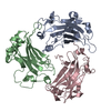


 PDBj
PDBj


 Assembly


 Mass: 150.087 Da / Num. of mol.: 1 / Source method: obtained synthetically / Formula: C4H6O6
 Mass: 106.120 Da / Num. of mol.: 1 / Source method: obtained synthetically / Formula: C4H10O3
 Mass: 18.015 Da / Num. of mol.: 136 / Source method: isolated from a natural source / Formula: H2O
 Sample preparation
 / Beamline: 21-ID-F / Wavelength: 0.97872 Å
 Processing