 Movie
Movie Controller


+ Open data

- Basic information
| Entry | Database: PDB / ID: 7d7s | ||||||
|---|---|---|---|---|---|---|---|
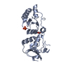
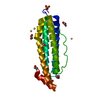
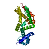
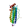
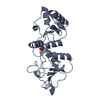
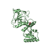
| Title | HIV-1 SF2 Nef in complex with the Fyn SH3 R96I mutant | ||||||
 Components Components |
| ||||||
 Keywords Keywords |  VIRAL PROTEIN / Complex / mutant / kinase / SH3 VIRAL PROTEIN / Complex / mutant / kinase / SH3 | ||||||
| Function / homology |  Function and homology information Function and homology informationsymbiont-mediated suppression of host antigen processing and presentation of peptide antigen via MHC class I / symbiont-mediated suppression of host antigen processing and presentation of peptide antigen via MHC class II / suppression by virus of host autophagy / activation of transmembrane receptor protein tyrosine kinase activity / host cell Golgi membrane / virion component / endocytosis involved in viral entry into host cell / SH3 domain binding / virus-mediated perturbation of host defense response / GTP binding ...symbiont-mediated suppression of host antigen processing and presentation of peptide antigen via MHC class I / symbiont-mediated suppression of host antigen processing and presentation of peptide antigen via MHC class II / suppression by virus of host autophagy / activation of transmembrane receptor protein tyrosine kinase activity / host cell Golgi membrane / virion component / endocytosis involved in viral entry into host cell / SH3 domain binding / virus-mediated perturbation of host defense response / GTP binding / host cell plasma membrane / extracellular region / membrane / plasma membrane / cytosolSimilarity search - Function | ||||||
| Biological species |  Human immunodeficiency virus type 1 group M subtype B Human immunodeficiency virus type 1 group M subtype B Homo sapiens (human) Homo sapiens (human) | ||||||
| Method | X-RAY DIFFRACTION / SYNCHROTRON / MOLECULAR REPLACEMENT / molecular replacement / Resolution: 3.32 Å | ||||||
 Authors Authors | Aldehaiman, A. / Shahul Hamed, U.F. / Arold, S.T. | ||||||
 Citation Citation | Journal: Biochem.J. / Year: 2021 Title: Synergy and allostery in ligand binding by HIV-1 Nef. Authors: Aldehaiman, A. / Momin, A.A. / Restouin, A. / Wang, L. / Shi, X. / Aljedani, S. / Opi, S. / Lugari, A. / Shahul Hameed, U.F. / Ponchon, L. / Morelli, X. / Huang, M. / Dumas, C. / Collette, Y. / Arold, S.T. | ||||||
| History |
|
- Structure visualization
| Structure viewer | Molecule: MolmilJmol/JSmol |
|---|
- Downloads & links
-Download
| PDBx/mmCIF format | 7d7s.cif.gz | 160.5 KB | Display | PDBx/mmCIF format |
|---|---|---|---|---|
| PDB format | pdb7d7s.ent.gz | 126.7 KB | Display | PDB format |
| PDBx/mmJSON format | 7d7s.json.gz | Tree view | PDBx/mmJSON format | |
| Others |  Other downloads Other downloads |
-Validation report
| Arichive directory | https://data.pdbj.org/pub/pdb/validation_reports/d7/7d7sftp://data.pdbj.org/pub/pdb/validation_reports/d7/7d7s | HTTPS FTP |
|---|
-Related structure data
| Related structure data |  3h0fC  3h0hC  3h0iC 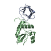 4d8dSC  6ipyC 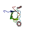 6ipzC S: Starting model for refinement C: citing same article ( |
|---|---|
| Similar structure data | |
| Experimental dataset #1 | Data reference: 10.25781/KAUST-8CV15 / Data set type: other data |


-Links
 PDBj
PDBj- Assembly
| Deposited unit | 
| ||||||||
|---|---|---|---|---|---|---|---|---|---|
| 1 | 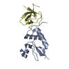
| ||||||||
| 2 | 
| ||||||||
| Unit cell |
|
-Components
| #1: Protein | Mass: 25585.838 Da / Num. of mol.: 2 Source method: isolated from a genetically manipulated source Source: (gene. exp.) Human immunodeficiency virus type 1 group M subtype B (isolate ARV2/SF2)Strain: isolate ARV2/SF2 / Gene: nef / Plasmid: pET23d / Production host:  Escherichia coli (E. coli) / References: UniProt: P03407 Escherichia coli (E. coli) / References: UniProt: P03407#2: Protein | Mass: 8254.987 Da / Num. of mol.: 2 / Mutation: R96I Source method: isolated from a genetically manipulated source Source: (gene. exp.) Homo sapiens (human) / Gene: FYN / Plasmid: pET42a / Production host: Escherichia coli (E. coli) / References: UniProt: E5RFS5 |
|---|
-Experimental details
-Experiment
| Experiment | Method: X-RAY DIFFRACTION / Number of used crystals: 3 |
|---|
- Sample preparation
| Crystal |
| ||||||||||||||||||||
|---|---|---|---|---|---|---|---|---|---|---|---|---|---|---|---|---|---|---|---|---|---|
| Crystal grow |
|
-Data collection
| Diffraction | Mean temperature: 100 K / Serial crystal experiment: N |
|---|---|
| Diffraction source | Source: SYNCHROTRON / Site: SOLEIL  / Beamline: PROXIMA 2 / Wavelength: 0.98011 Å / Beamline: PROXIMA 2 / Wavelength: 0.98011 Å |
| Detector | Type: DECTRIS EIGER2 X 9M / Detector: PIXEL / Date: Feb 11, 2020 Details: focusing bimorph mirrors in Kirkpatrick-Baez (KB) configuration |
| Radiation | Monochromator: cryogenically cooled channel-cut Si[111] / Protocol: SINGLE WAVELENGTH / Monochromatic (M) / Laue (L): M / Scattering type: x-ray |
| Radiation wavelength | Wavelength: 0.98011 Å / Relative weight: 1 |
| Reflection | Resolution: 3.32→48.68 Å / Num. obs: 9209 / % possible obs: 94.6 % / Redundancy: 155.4 % / CC1/2: 1 / Rmerge(I) obs: 0.218 / Rpim(I) all: 0.018 / Rrim(I) all: 0.219 / Net I/σ(I): 33.6 |
| Reflection shell | Resolution: 3.324→3.46 Å / Redundancy: 165.4 % / Rmerge(I) obs: 9.136 / Mean I/σ(I) obs: 1.1 / Num. unique obs: 461 / CC1/2: 0.486 / Rpim(I) all: 0.71 / Rrim(I) all: 9.163 / % possible all: 42.1 |
-Phasing
| Phasing | Method: molecular replacement |
|---|
- Processing
| Software |
| ||||||||||||||||||||||||||||||||||||||||||||||||||||||||||||||||||||||||||||||||||||||||||||||||||||||||||||||||||||||||||||||||||||||||||||||||||||||||||||||||||||||||||||||||||||||
|---|---|---|---|---|---|---|---|---|---|---|---|---|---|---|---|---|---|---|---|---|---|---|---|---|---|---|---|---|---|---|---|---|---|---|---|---|---|---|---|---|---|---|---|---|---|---|---|---|---|---|---|---|---|---|---|---|---|---|---|---|---|---|---|---|---|---|---|---|---|---|---|---|---|---|---|---|---|---|---|---|---|---|---|---|---|---|---|---|---|---|---|---|---|---|---|---|---|---|---|---|---|---|---|---|---|---|---|---|---|---|---|---|---|---|---|---|---|---|---|---|---|---|---|---|---|---|---|---|---|---|---|---|---|---|---|---|---|---|---|---|---|---|---|---|---|---|---|---|---|---|---|---|---|---|---|---|---|---|---|---|---|---|---|---|---|---|---|---|---|---|---|---|---|---|---|---|---|---|---|---|---|---|---|
| Refinement | Method to determine structure: MOLECULAR REPLACEMENT Starting model: 4d8d Resolution: 3.32→48.68 Å / Cor.coef. Fo:Fc: 0.896 / Cor.coef. Fo:Fc free: 0.9 / SU B: 109.02 / SU ML: 0.715 / Cross valid method: THROUGHOUT / ESU R: 6.24 / ESU R Free: 0.609 / Stereochemistry target values: MAXIMUM LIKELIHOOD / Details: HYDROGENS HAVE BEEN ADDED IN THE RIDING POSITIONS
| ||||||||||||||||||||||||||||||||||||||||||||||||||||||||||||||||||||||||||||||||||||||||||||||||||||||||||||||||||||||||||||||||||||||||||||||||||||||||||||||||||||||||||||||||||||||
| Solvent computation | Ion probe radii: 0.7 Å / Shrinkage radii: 0.7 Å / VDW probe radii: 1.1 Å / Solvent model: MASK | ||||||||||||||||||||||||||||||||||||||||||||||||||||||||||||||||||||||||||||||||||||||||||||||||||||||||||||||||||||||||||||||||||||||||||||||||||||||||||||||||||||||||||||||||||||||
| Displacement parameters | Biso mean: 150.582 Å2
| ||||||||||||||||||||||||||||||||||||||||||||||||||||||||||||||||||||||||||||||||||||||||||||||||||||||||||||||||||||||||||||||||||||||||||||||||||||||||||||||||||||||||||||||||||||||
| Refinement step | Cycle: LAST / Resolution: 3.32→48.68 Å
| ||||||||||||||||||||||||||||||||||||||||||||||||||||||||||||||||||||||||||||||||||||||||||||||||||||||||||||||||||||||||||||||||||||||||||||||||||||||||||||||||||||||||||||||||||||||
| Refine LS restraints |
|