 Movie
Movie Controller

[English] 日本語
 Yorodumi
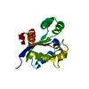
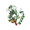
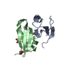
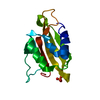
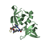
- PDB-3llo: Crystal structure of the STAS domain of motor protein prestin (an... -
+ Open data

- Basic information
| Entry | Database: PDB / ID: 3llo | ||||||
|---|---|---|---|---|---|---|---|
| Title | Crystal structure of the STAS domain of motor protein prestin (anion transporter SLC26A5) | ||||||
 Components Components | Prestin | ||||||
 Keywords Keywords | MOTOR PROTEIN / STAS domain / Cell shape / Glycoprotein / Membrane / Transmembrane | ||||||
| Function / homology |  Function and homology information Function and homology informationoxalate transport / lateral wall of outer hair cell / fructose transmembrane transport / response to salicylic acid / sulfate transmembrane transporter activity / negative regulation of monoatomic ion transmembrane transport / secondary active sulfate transmembrane transporter activity / oxalate transmembrane transporter activity / response to potassium ion / response to auditory stimulus ...oxalate transport / lateral wall of outer hair cell / fructose transmembrane transport / response to salicylic acid / sulfate transmembrane transporter activity / negative regulation of monoatomic ion transmembrane transport / secondary active sulfate transmembrane transporter activity / oxalate transmembrane transporter activity / response to potassium ion / response to auditory stimulus / monoatomic anion transmembrane transport / response to salt / chloride:bicarbonate antiporter activity / response to thyroid hormone / bicarbonate transport / bicarbonate transmembrane transporter activity / positive regulation of cell motility / chloride transport / chloride transmembrane transporter activity / spectrin binding / cochlea development / cytoskeletal motor activity / positive regulation of cell size / lateral plasma membrane / monoatomic ion transmembrane transport / chloride transmembrane transport / regulation of membrane potential / response to ischemia / sensory perception of sound / regulation of cell shape / basolateral plasma membrane / response to xenobiotic stimulus / protein homodimerization activity / identical protein binding / plasma membraneSimilarity search - Function | ||||||
| Biological species |  Rattus norvegicus (Norway rat) Rattus norvegicus (Norway rat) | ||||||
| Method | X-RAY DIFFRACTION / SYNCHROTRON / SAD / molecular replacement / Resolution: 1.57 Å | ||||||
 Authors Authors | Pasqualetto, E. / Aiello, R. / Bonetto, G. / Battistutta, R. | ||||||
 Citation Citation | Journal: J.Mol.Biol. / Year: 2010 Title: Structure of the cytosolic portion of the motor protein prestin and functional role of the STAS domain in SLC26/SulP anion transporters. Authors: Pasqualetto, E. / Aiello, R. / Gesiot, L. / Bonetto, G. / Bellanda, M. / Battistutta, R. | ||||||
| History |
|
- Structure visualization
| Structure viewer | Molecule: MolmilJmol/JSmol |
|---|
- Downloads & links
-Download
| PDBx/mmCIF format | 3llo.cif.gz | 71.5 KB | Display | PDBx/mmCIF format |
|---|---|---|---|---|
| PDB format | pdb3llo.ent.gz | 52 KB | Display | PDB format |
| PDBx/mmJSON format | 3llo.json.gz | Tree view | PDBx/mmJSON format | |
| Others |  Other downloads Other downloads |
-Validation report
| Arichive directory | https://data.pdbj.org/pub/pdb/validation_reports/ll/3lloftp://data.pdbj.org/pub/pdb/validation_reports/ll/3llo | HTTPS FTP |
|---|
-Related structure data
| Similar structure data |
|---|


-Links
 PDBj
PDBj
- Assembly
| Deposited unit | 
| ||||||||
|---|---|---|---|---|---|---|---|---|---|
| 1 |
| ||||||||
| Unit cell |
|
-Components
| #1: Protein | / Solute carrier family 26 member 5 Mass: 15766.707 Da / Num. of mol.: 1 / Fragment: UNP residues 505-563, 637-718 Source method: isolated from a genetically manipulated source Source: (gene. exp.) Rattus norvegicus (Norway rat) / Gene: Slc26a5, Pres / Production host:  Escherichia coli (E. coli) / References: UniProt: Q9EPH0 Escherichia coli (E. coli) / References: UniProt: Q9EPH0 |
|---|---|
| #2: Sugar | ChemComp-BOG / Octyl glucoside  Type: D-saccharide / Mass: 292.369 Da / Num. of mol.: 1 Type: D-saccharide / Mass: 292.369 Da / Num. of mol.: 1Source method: isolated from a genetically manipulated source Formula: C14H28O6 / Comment: detergent*YM |
| #3: Water | ChemComp-HOH / Water Mass: 18.015 Da / Num. of mol.: 103 / Source method: isolated from a natural source / Formula: H2O Mass: 18.015 Da / Num. of mol.: 103 / Source method: isolated from a natural source / Formula: H2O |
-Experimental details
-Experiment
| Experiment | Method: X-RAY DIFFRACTION / Number of used crystals: 1 |
|---|
- Sample preparation
| Crystal | Density Matthews: 2.33 Å3/Da / Density % sol: 47.24 % Description: The Structure Factor File contains Friedel Pairs |
|---|---|
| Crystal grow | Temperature: 293 K / Method: vapor diffusion / pH: 6.5 Details: 1.8 M Ammonium sulfate, 4.5% (v/v) PEG 400, 0.1% (w/v) octyl-beta-D-glucopyranoside, 0.09 M MES, pH 6.5, VAPOR DIFFUSION, temperature 293.0K |
-Data collection
| Diffraction | Mean temperature: 100 K | |||||||||||||||||||||||||||||||||||||||||||||||||||||||||||||||||||||||||||||
|---|---|---|---|---|---|---|---|---|---|---|---|---|---|---|---|---|---|---|---|---|---|---|---|---|---|---|---|---|---|---|---|---|---|---|---|---|---|---|---|---|---|---|---|---|---|---|---|---|---|---|---|---|---|---|---|---|---|---|---|---|---|---|---|---|---|---|---|---|---|---|---|---|---|---|---|---|---|---|
| Diffraction source | Source: SYNCHROTRON / Site: ESRF  / Beamline: ID14-1 / Wavelength: 0.93 Å / Beamline: ID14-1 / Wavelength: 0.93 Å | |||||||||||||||||||||||||||||||||||||||||||||||||||||||||||||||||||||||||||||
| Detector | Type: ADSC QUANTUM 210 / Detector: CCD / Date: Sep 8, 2009 | |||||||||||||||||||||||||||||||||||||||||||||||||||||||||||||||||||||||||||||
| Radiation | Monochromator: Diamond (111) / Protocol: SINGLE WAVELENGTH / Monochromatic (M) / Laue (L): M / Scattering type: x-ray | |||||||||||||||||||||||||||||||||||||||||||||||||||||||||||||||||||||||||||||
| Radiation wavelength | Wavelength: 0.93 Å / Relative weight: 1 | |||||||||||||||||||||||||||||||||||||||||||||||||||||||||||||||||||||||||||||
| Reflection | Redundancy: 32.7 % / Av σ(I) over netI: 6.5 / Number: 660833 / Rsym value: 0.066 / D res high: 1.6 Å / D res low: 53.64 Å / Num. obs: 20184 / % possible obs: 100 | |||||||||||||||||||||||||||||||||||||||||||||||||||||||||||||||||||||||||||||
| Diffraction reflection shell |
| |||||||||||||||||||||||||||||||||||||||||||||||||||||||||||||||||||||||||||||
| Reflection | Resolution: 1.57→28.408 Å / Num. all: 20860 / Num. obs: 20860 / % possible obs: 99.3 % / Redundancy: 11.3 % / Biso Wilson estimate: 21.4 Å2 / Rmerge(I) obs: 0.055 / Rsym value: 0.055 / Net I/σ(I): 8.5 | |||||||||||||||||||||||||||||||||||||||||||||||||||||||||||||||||||||||||||||
| Reflection shell | Resolution: 1.57→1.65 Å / Redundancy: 10.1 % / Rmerge(I) obs: 0.496 / Mean I/σ(I) obs: 3.9 / Num. unique all: 2931 / Rsym value: 0.496 / % possible all: 97.6 |
-Phasing
| Phasing |
| ||||||||||||||||||||||||||||||||||||||||||||||||||||||||||||||||||||||||||||||||||||||||||||||||||||||||||||||||
|---|---|---|---|---|---|---|---|---|---|---|---|---|---|---|---|---|---|---|---|---|---|---|---|---|---|---|---|---|---|---|---|---|---|---|---|---|---|---|---|---|---|---|---|---|---|---|---|---|---|---|---|---|---|---|---|---|---|---|---|---|---|---|---|---|---|---|---|---|---|---|---|---|---|---|---|---|---|---|---|---|---|---|---|---|---|---|---|---|---|---|---|---|---|---|---|---|---|---|---|---|---|---|---|---|---|---|---|---|---|---|---|---|---|
| Phasing MR | Rfactor: 28.82 / Model details: Phaser MODE: MR_AUTO
| ||||||||||||||||||||||||||||||||||||||||||||||||||||||||||||||||||||||||||||||||||||||||||||||||||||||||||||||||
| Phasing dm | Method: Solvent flattening and Histogram matching / Reflection: 20160 | ||||||||||||||||||||||||||||||||||||||||||||||||||||||||||||||||||||||||||||||||||||||||||||||||||||||||||||||||
| Phasing dm shell |
|
- Processing
| Software |
| ||||||||||||||||||||||||||||||||||||||||||||||||||||||||||||||||||||||
|---|---|---|---|---|---|---|---|---|---|---|---|---|---|---|---|---|---|---|---|---|---|---|---|---|---|---|---|---|---|---|---|---|---|---|---|---|---|---|---|---|---|---|---|---|---|---|---|---|---|---|---|---|---|---|---|---|---|---|---|---|---|---|---|---|---|---|---|---|---|---|---|
| Refinement | Method to determine structure: SAD / Resolution: 1.57→25 Å / Cor.coef. Fo:Fc: 0.968 / Cor.coef. Fo:Fc free: 0.958 / Occupancy max: 1 / Occupancy min: 0.5 / SU B: 2.677 / SU ML: 0.044 / Cross valid method: THROUGHOUT / σ(F): 0 / ESU R: 0.087 / ESU R Free: 0.076 / Stereochemistry target values: MAXIMUM LIKELIHOOD Details: HYDROGENS HAVE BEEN ADDED IN THE RIDING POSITIONS. U VALUES: REFINED INDIVIDUALLY
| ||||||||||||||||||||||||||||||||||||||||||||||||||||||||||||||||||||||
| Solvent computation | Ion probe radii: 0.8 Å / Shrinkage radii: 0.8 Å / VDW probe radii: 1.4 Å / Solvent model: MASK | ||||||||||||||||||||||||||||||||||||||||||||||||||||||||||||||||||||||
| Displacement parameters | Biso max: 70.27 Å2 / Biso mean: 25.149 Å2 / Biso min: 11.26 Å2
| ||||||||||||||||||||||||||||||||||||||||||||||||||||||||||||||||||||||
| Refinement step | Cycle: LAST / Resolution: 1.57→25 Å
| ||||||||||||||||||||||||||||||||||||||||||||||||||||||||||||||||||||||
| Refine LS restraints |
| ||||||||||||||||||||||||||||||||||||||||||||||||||||||||||||||||||||||
| LS refinement shell | Resolution: 1.57→1.611 Å / Total num. of bins used: 20
|
