 Movie
Movie Controller

[English] 日本語
 Yorodumi
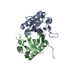
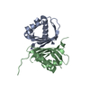
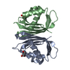
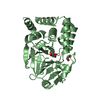
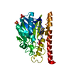
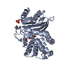
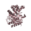
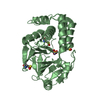
- PDB-4iia: Low resolution crystal structure of the NTF2-like domain of human... -
+ Open data

- Basic information
| Entry | Database: PDB / ID: 4iia | ||||||
|---|---|---|---|---|---|---|---|
| Title | Low resolution crystal structure of the NTF2-like domain of human G3BP1 | ||||||
 Components Components | Ras GTPase-activating protein-binding protein 1 | ||||||
 Keywords Keywords |  HYDROLASE / NTF2-LIKE DOMAIN HYDROLASE / NTF2-LIKE DOMAIN | ||||||
| Function / homology |  Function and homology information Function and homology informationDNA/RNA helicase activity / positive regulation of stress granule assembly / ribosomal small subunit binding / positive regulation of type I interferon production / stress granule assembly / DNA helicase activity / molecular condensate scaffold activity / negative regulation of canonical Wnt signaling pathway / cytoplasmic stress granule / perikaryon ...DNA/RNA helicase activity / positive regulation of stress granule assembly / ribosomal small subunit binding / positive regulation of type I interferon production / stress granule assembly / DNA helicase activity / molecular condensate scaffold activity / negative regulation of canonical Wnt signaling pathway / cytoplasmic stress granule / perikaryon / endonuclease activity / defense response to virus / DNA helicase / Ras protein signal transduction / RNA helicase activity / RNA helicase / ribonucleoprotein complex / focal adhesion / innate immune response / mRNA binding / SARS-CoV-2 activates/modulates innate and adaptive immune responses / ATP hydrolysis activity / DNA binding / RNA binding / ATP binding / nucleus / cytosol / cytoplasmSimilarity search - Function | ||||||
| Biological species |  Homo sapiens (human) Homo sapiens (human) | ||||||
| Method | X-RAY DIFFRACTION / SYNCHROTRON / SIRAS, molecular replacement / Resolution: 3.3 Å | ||||||
 Authors Authors | Vognsen, T. / Moeller, I.R. / Kristensen, O. | ||||||
 Citation Citation | Journal: Plos One / Year: 2013 Title: Crystal Structures of the Human G3BP1 NTF2-Like Domain Visualize FxFG Nup Repeat Specificity. Authors: Vognsen, T. / Moller, I.R. / Kristensen, O. #1: Journal: Acta Crystallogr.,Sect.F / Year: 2011 Title: Purification, crystallization and preliminary X-ray diffraction of the G3BP1 NTF2-like domain. Authors: Vognsen, T. / Moller, I.R. / Kristensen, O. #2: Journal: Biochem.Biophys.Res.Commun. / Year: 2012Title: Crystal structure of the Rasputin NTF2-like domain from Drosophila melanogaster. Authors: Vognsen, T. / Kristensen, O. | ||||||
| History |
|
- Structure visualization
| Structure viewer | Molecule: MolmilJmol/JSmol |
|---|
- Downloads & links
-Download
| PDBx/mmCIF format | 4iia.cif.gz | 39.3 KB | Display | PDBx/mmCIF format |
|---|---|---|---|---|
| PDB format | pdb4iia.ent.gz | 27.7 KB | Display | PDB format |
| PDBx/mmJSON format | 4iia.json.gz | Tree view | PDBx/mmJSON format | |
| Others |  Other downloads Other downloads |
-Validation report
| Arichive directory | https://data.pdbj.org/pub/pdb/validation_reports/ii/4iiaftp://data.pdbj.org/pub/pdb/validation_reports/ii/4iia | HTTPS FTP |
|---|
-Related structure data


-Links
 PDBj
PDBj
- Assembly
| Deposited unit | 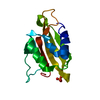
| ||||||||||||
|---|---|---|---|---|---|---|---|---|---|---|---|---|---|
| 1 | 
| ||||||||||||
| Unit cell |
| ||||||||||||
| Components on special symmetry positions |
|

-Components
| #1: Protein | Mass: 15185.106 Da / Num. of mol.: 1 / Fragment: NTF2-LIKE DOMAIN Source method: isolated from a genetically manipulated source Source: (gene. exp.) Homo sapiens (human) / Gene: G3BP1, G3BP / Production host:  Escherichia coli (E. coli) / References: UniProt: Q13283, DNA helicase, RNA helicase Escherichia coli (E. coli) / References: UniProt: Q13283, DNA helicase, RNA helicase |
|---|---|
| #2: Chemical | ChemComp-PO4 / Phosphate  Mass: 94.971 Da / Num. of mol.: 1 / Source method: obtained synthetically / Formula: PO4 Mass: 94.971 Da / Num. of mol.: 1 / Source method: obtained synthetically / Formula: PO4 |
-Experimental details
-Experiment
| Experiment | Method: X-RAY DIFFRACTION / Number of used crystals: 1 |
|---|
- Sample preparation
| Crystal | Density Matthews: 2.66 Å3/Da / Density % sol: 53.83 % |
|---|---|
| Crystal grow | Temperature: 293 K / Method: vapor diffusion, hanging drop / pH: 8 Details: 1.6 M diammonium phosphate, 0.1 M MOPS, pH 8.0, VAPOR DIFFUSION, HANGING DROP, temperature 293K |
-Data collection
| Diffraction | Mean temperature: 100 K |
|---|---|
| Diffraction source | Source: SYNCHROTRON / Site: MAX II  / Beamline: I911-2 / Wavelength: 1.04 Å / Beamline: I911-2 / Wavelength: 1.04 Å |
| Detector | Type: MAR CCD 165 mm / Detector: CCD / Date: May 27, 2010 |
| Radiation | Monochromator: BENT SI (111) CRYSTAL / Protocol: SINGLE WAVELENGTH / Monochromatic (M) / Laue (L): M / Scattering type: x-ray |
| Radiation wavelength | Wavelength: 1.04 Å / Relative weight: 1 |
| Reflection | Resolution: 3.3→29.35 Å / Num. obs: 2719 / % possible obs: 98.7 % / Observed criterion σ(F): 0 / Observed criterion σ(I): 0 / Redundancy: 10 % / Biso Wilson estimate: 121.6 Å2 / Rmerge(I) obs: 0.085 / Rsym value: 0.085 / Net I/σ(I): 15.7 |
| Reflection shell | Resolution: 3.3→3.39 Å / Redundancy: 10.1 % / Mean I/σ(I) obs: 1.5 / % possible all: 99.1 |
- Processing
| Software |
| ||||||||||||||||||||||||||||
|---|---|---|---|---|---|---|---|---|---|---|---|---|---|---|---|---|---|---|---|---|---|---|---|---|---|---|---|---|---|
| Refinement | Method to determine structure: SIRAS, molecular replacement / Resolution: 3.3→27.006 Å / SU ML: 0.3 / σ(F): 1.35 / Phase error: 48 / Stereochemistry target values: MLHL
| ||||||||||||||||||||||||||||
| Solvent computation | Shrinkage radii: 0.9 Å / VDW probe radii: 1.11 Å / Solvent model: FLAT BULK SOLVENT MODEL | ||||||||||||||||||||||||||||
| Refinement step | Cycle: LAST / Resolution: 3.3→27.006 Å
| ||||||||||||||||||||||||||||
| Refine LS restraints |
| ||||||||||||||||||||||||||||
| LS refinement shell |
|
