 Movie
Movie Controller

[English] 日本語
 Yorodumi
- PDB-3q90: Crystal structure of the NTF2 domain of Ras GTPase-activating pro... -
+ Open data

- Basic information
| Entry | Database: PDB / ID: 3q90 | ||||||
|---|---|---|---|---|---|---|---|
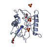
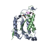
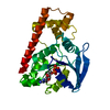
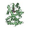
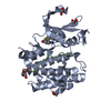
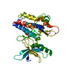
| Title | Crystal structure of the NTF2 domain of Ras GTPase-activating protein-binding protein 1 | ||||||
 Components Components | Ras GTPase-activating protein-binding protein 1 | ||||||
 Keywords Keywords |  HYDROLASE / Structural Genomics / Structural Genomics Consortium / SGC / NTF2-like (a+b proteins) / Protein Binding and Helicase / Protein (Ras GTPase-activating protein) / DNA and RNA binding / plasma membrane / nucleus HYDROLASE / Structural Genomics / Structural Genomics Consortium / SGC / NTF2-like (a+b proteins) / Protein Binding and Helicase / Protein (Ras GTPase-activating protein) / DNA and RNA binding / plasma membrane / nucleus | ||||||
| Function / homology |  Function and homology information Function and homology informationDNA/RNA helicase activity / positive regulation of stress granule assembly / ribosomal small subunit binding / positive regulation of type I interferon production / stress granule assembly / DNA helicase activity / molecular condensate scaffold activity / negative regulation of canonical Wnt signaling pathway / cytoplasmic stress granule / perikaryon ...DNA/RNA helicase activity / positive regulation of stress granule assembly / ribosomal small subunit binding / positive regulation of type I interferon production / stress granule assembly / DNA helicase activity / molecular condensate scaffold activity / negative regulation of canonical Wnt signaling pathway / cytoplasmic stress granule / perikaryon / endonuclease activity / defense response to virus / DNA helicase / Ras protein signal transduction / RNA helicase activity / RNA helicase / ribonucleoprotein complex / focal adhesion / innate immune response / mRNA binding / SARS-CoV-2 activates/modulates innate and adaptive immune responses / ATP hydrolysis activity / DNA binding / RNA binding / ATP binding / nucleus / cytosol / cytoplasmSimilarity search - Function | ||||||
| Biological species |  Homo sapiens (human) Homo sapiens (human) | ||||||
| Method | X-RAY DIFFRACTION / SYNCHROTRON / MOLECULAR REPLACEMENT / Resolution: 1.7 Å | ||||||
 Authors Authors | Welin, M. / Tresaugues, L. / Arrowsmith, C.H. / Berglund, H. / Bountra, C. / Collins, R. / Edwards, A.M. / Ekblad, T. / Flodin, S. / Flores, A. ...Welin, M. / Tresaugues, L. / Arrowsmith, C.H. / Berglund, H. / Bountra, C. / Collins, R. / Edwards, A.M. / Ekblad, T. / Flodin, S. / Flores, A. / Graslund, S. / Hammarstrom, M. / Johansson, I. / Karlberg, T. / Kol, S. / Kotenyova, T. / Kouznetsova, E. / Moche, M. / Nyman, T. / Persson, C. / Schuler, H. / Schutz, P. / Siponen, M.I. / Thorsell, A.G. / Van Der Berg, S. / Wahlberg, E. / Weigelt, J. / Nordlund, P. / Structural Genomics Consortium (SGC) | ||||||
 Citation Citation | Journal: To be Published Title: Crystal structure of the NTF2 domain of Ras GTPase-activating protein-binding protein 1 Authors: Welin, M. / Tresaugues, L. / Arrowsmith, C.H. / Berglund, H. / Bountra, C. / Collins, R. / Edwards, A.M. / Ekblad, T. / Flodin, S. / Flores, A. / Graslund, S. / Hammarstrom, M. / Johansson, ...Authors: Welin, M. / Tresaugues, L. / Arrowsmith, C.H. / Berglund, H. / Bountra, C. / Collins, R. / Edwards, A.M. / Ekblad, T. / Flodin, S. / Flores, A. / Graslund, S. / Hammarstrom, M. / Johansson, I. / Karlberg, T. / Kol, S. / Kotenyova, T. / Kouznetsova, E. / Moche, M. / Nyman, T. / Persson, C. / Schuler, H. / Schutz, P. / Siponen, M.I. / Thorsell, A.G. / Van Der Berg, S. / Wahlberg, E. / Weigelt, J. / Nordlund, P. | ||||||
| History |
|
- Structure visualization
| Structure viewer | Molecule: MolmilJmol/JSmol |
|---|
- Downloads & links
-Download
| PDBx/mmCIF format | 3q90.cif.gz | 115.8 KB | Display | PDBx/mmCIF format |
|---|---|---|---|---|
| PDB format | pdb3q90.ent.gz | 91.2 KB | Display | PDB format |
| PDBx/mmJSON format | 3q90.json.gz | Tree view | PDBx/mmJSON format | |
| Others |  Other downloads Other downloads |
-Validation report
| Arichive directory | https://data.pdbj.org/pub/pdb/validation_reports/q9/3q90ftp://data.pdbj.org/pub/pdb/validation_reports/q9/3q90 | HTTPS FTP |
|---|
-Related structure data
| Related structure data |  1z02S S: Starting model for refinement |
|---|---|
| Similar structure data |


-Links
 PDBj
PDBj
- Assembly
| Deposited unit | 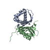
| ||||||||
|---|---|---|---|---|---|---|---|---|---|
| 1 |
| ||||||||
| Unit cell |
|
-Components
| #1: Protein | Mass: 15986.153 Da / Num. of mol.: 2 / Fragment: unp residues 1-139 Source method: isolated from a genetically manipulated source Source: (gene. exp.) Homo sapiens (human) / Gene: G3BP, G3BP1 / Plasmid: pNIC-Bsa4 / Production host:  Escherichia coli (E. coli) / Strain (production host): BL21(DE3) R3 pRARE / References: UniProt: Q13283, DNA helicase, RNA helicase Escherichia coli (E. coli) / Strain (production host): BL21(DE3) R3 pRARE / References: UniProt: Q13283, DNA helicase, RNA helicase#2: Water | ChemComp-HOH / | Water Mass: 18.015 Da / Num. of mol.: 119 / Source method: isolated from a natural source / Formula: H2O Mass: 18.015 Da / Num. of mol.: 119 / Source method: isolated from a natural source / Formula: H2O |
|---|
-Experimental details
-Experiment
| Experiment | Method: X-RAY DIFFRACTION / Number of used crystals: 1 |
|---|
- Sample preparation
| Crystal | Density Matthews: 2.12 Å3/Da / Density % sol: 41.97 % |
|---|---|
| Crystal grow | Temperature: 277 K / Method: vapor diffusion, sitting drop / pH: 5.5 Details: 25% w/v PEG3350, 0.2M AMMONIUM ACETATE, 0.1M BIS-TRIS, VAPOR DIFFUSION, SITTING DROP, temperature 277K, pH 5.5 |
-Data collection
| Diffraction | Mean temperature: 100 K |
|---|---|
| Diffraction source | Source: SYNCHROTRON / Site: ESRF  / Beamline: ID23-2 / Wavelength: 0.8726 Å / Beamline: ID23-2 / Wavelength: 0.8726 Å |
| Detector | Type: MARMOSAIC 225 mm CCD / Detector: CCD / Date: Jun 20, 2010 / Details: MIRRORS |
| Radiation | Monochromator: horizontally side diffracting Silicon 111 crystal Protocol: SINGLE WAVELENGTH / Monochromatic (M) / Laue (L): M / Scattering type: x-ray |
| Radiation wavelength | Wavelength: 0.8726 Å / Relative weight: 1 |
| Reflection | Resolution: 1.7→50 Å / Num. all: 30636 / Num. obs: 30582 / % possible obs: 99.8 % / Observed criterion σ(I): -3 / Redundancy: 6.17 % / Biso Wilson estimate: 22.39 Å2 / Rsym value: 0.063 / Net I/σ(I): 17.17 |
| Reflection shell | Resolution: 1.7→1.8 Å / Redundancy: 3.67 % / Mean I/σ(I) obs: 3.48 / Num. unique all: 4585 / Rsym value: 0.434 / % possible all: 99.8 |
- Processing
| Software |
| |||||||||||||||||||||||||||||||||||||||||||||||||||||||||||||||||||||||||||
|---|---|---|---|---|---|---|---|---|---|---|---|---|---|---|---|---|---|---|---|---|---|---|---|---|---|---|---|---|---|---|---|---|---|---|---|---|---|---|---|---|---|---|---|---|---|---|---|---|---|---|---|---|---|---|---|---|---|---|---|---|---|---|---|---|---|---|---|---|---|---|---|---|---|---|---|---|
| Refinement | Method to determine structure: MOLECULAR REPLACEMENT Starting model: PDB ENTRY 1Z02 Resolution: 1.7→44.29 Å / Cor.coef. Fo:Fc: 0.9302 / Cor.coef. Fo:Fc free: 0.9175 / Cross valid method: THROUGHOUT / σ(F): 0
| |||||||||||||||||||||||||||||||||||||||||||||||||||||||||||||||||||||||||||
| Displacement parameters | Biso mean: 29.55 Å2
| |||||||||||||||||||||||||||||||||||||||||||||||||||||||||||||||||||||||||||
| Refine analyze | Luzzati coordinate error obs: 0.239 Å | |||||||||||||||||||||||||||||||||||||||||||||||||||||||||||||||||||||||||||
| Refinement step | Cycle: LAST / Resolution: 1.7→44.29 Å
| |||||||||||||||||||||||||||||||||||||||||||||||||||||||||||||||||||||||||||
| Refine LS restraints |
| |||||||||||||||||||||||||||||||||||||||||||||||||||||||||||||||||||||||||||
| LS refinement shell | Resolution: 1.7→1.76 Å / Total num. of bins used: 15
| |||||||||||||||||||||||||||||||||||||||||||||||||||||||||||||||||||||||||||
| Refinement TLS params. | Method: refined / Refine-ID: X-RAY DIFFRACTION
| |||||||||||||||||||||||||||||||||||||||||||||||||||||||||||||||||||||||||||
| Refinement TLS group |
|
