 Movie
Movie Controller


+ Open data

- Basic information
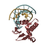
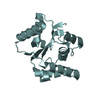
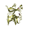
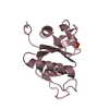
| Entry | Database: PDB / ID: 5euu | |||||||||
|---|---|---|---|---|---|---|---|---|---|---|
| Title | Rat prestin STAS domain in complex with chloride | |||||||||
 Components Components | Prestin,Prestin | |||||||||
 Keywords Keywords |  TRANSPORT PROTEIN / Anion-binding site / protein-anion complex TRANSPORT PROTEIN / Anion-binding site / protein-anion complex | |||||||||
| Function / homology |  Function and homology information Function and homology informationoxalate transport / lateral wall of outer hair cell / fructose transmembrane transport / response to salicylic acid / sulfate transmembrane transporter activity / negative regulation of monoatomic ion transmembrane transport / secondary active sulfate transmembrane transporter activity / oxalate transmembrane transporter activity / response to potassium ion / response to auditory stimulus ...oxalate transport / lateral wall of outer hair cell / fructose transmembrane transport / response to salicylic acid / sulfate transmembrane transporter activity / negative regulation of monoatomic ion transmembrane transport / secondary active sulfate transmembrane transporter activity / oxalate transmembrane transporter activity / response to potassium ion / response to auditory stimulus / monoatomic anion transmembrane transport / response to salt / chloride:bicarbonate antiporter activity / response to thyroid hormone / bicarbonate transport / bicarbonate transmembrane transporter activity / positive regulation of cell motility / chloride transport / chloride transmembrane transporter activity / spectrin binding / cochlea development / cytoskeletal motor activity / positive regulation of cell size / lateral plasma membrane / monoatomic ion transmembrane transport / chloride transmembrane transport / regulation of membrane potential / response to ischemia / sensory perception of sound / regulation of cell shape / basolateral plasma membrane / response to xenobiotic stimulus / protein homodimerization activity / identical protein binding / plasma membraneSimilarity search - Function | |||||||||
| Biological species |  Rattus norvegicus (Norway rat) Rattus norvegicus (Norway rat) | |||||||||
| Method | X-RAY DIFFRACTION / SYNCHROTRON / MOLECULAR REPLACEMENT / Resolution: 1.87 Å | |||||||||
 Authors Authors | Lolli, G. / Pasqualetto, E. / Costanzi, E. / Bonetto, G. / Battistutta, R. | |||||||||
| Funding support |  Italy, 2items Italy, 2items
| |||||||||
 Citation Citation | Journal: Biochem.J. / Year: 2016 Title: The STAS domain of mammalian SLC26A5 prestin harbours an anion-binding site. Authors: Lolli, G. / Pasqualetto, E. / Costanzi, E. / Bonetto, G. / Battistutta, R. #1: Journal: J.Mol.Biol. / Year: 2010Title: Structure of the Cytosolic Portion of the Motor Protein Prestin and Functional Role of the STAS Domain in SLC26/SulP Anion Transporters Authors: Pasqualetto, E. / Aiello, R. / Gesiot, L. / Bonetto, G. / Bellanda, M. / Battistutta, R. | |||||||||
| History |
|
- Structure visualization
| Structure viewer | Molecule: MolmilJmol/JSmol |
|---|
- Downloads & links
-Download
| PDBx/mmCIF format | 5euu.cif.gz | 92.4 KB | Display | PDBx/mmCIF format |
|---|---|---|---|---|
| PDB format | pdb5euu.ent.gz | 70.8 KB | Display | PDB format |
| PDBx/mmJSON format | 5euu.json.gz | Tree view | PDBx/mmJSON format | |
| Others |  Other downloads Other downloads |
-Validation report
| Arichive directory | https://data.pdbj.org/pub/pdb/validation_reports/eu/5euuftp://data.pdbj.org/pub/pdb/validation_reports/eu/5euu | HTTPS FTP |
|---|
-Related structure data
| Related structure data | 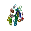 5eusC 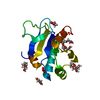 5euwC 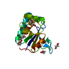 5euxC  5euzC  5ezbC 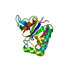 3lloS S: Starting model for refinement C: citing same article ( |
|---|---|
| Similar structure data |


-Links
 PDBj
PDBj
- Assembly
| Deposited unit | 
| ||||||||
|---|---|---|---|---|---|---|---|---|---|
| 1 |
| ||||||||
| Unit cell |
|
-Components
-Protein , 1 types, 1 molecules A
| #1: Protein | Mass: 15766.707 Da / Num. of mol.: 1 / Fragment: STAS domain,STAS domain,STAS domain,STAS domain Mutation: Residues 564-636 (variable loop) are deleted, GlySer are inserted between position 563 and 637,Residues 564-636 (variable loop) are deleted, GlySer are inserted between position 563 and ...Mutation: Residues 564-636 (variable loop) are deleted, GlySer are inserted between position 563 and 637,Residues 564-636 (variable loop) are deleted, GlySer are inserted between position 563 and 637,Residues 564-636 (variable loop) are deleted, GlySer are inserted between position 563 and 637,Residues 564-636 (variable loop) are deleted, GlySer are inserted between position 563 and 637 Source method: isolated from a genetically manipulated source Details: Residues 564-636 (variable loop) are deleted, GlySer are inserted between position 563 and 637 Source: (gene. exp.) Rattus norvegicus (Norway rat) / Gene: Slc26a5, Pres / Plasmid: pET SUMO / Production host:  Escherichia coli BL21(DE3) (bacteria) / References: UniProt: Q9EPH0 Escherichia coli BL21(DE3) (bacteria) / References: UniProt: Q9EPH0 |
|---|
-Non-polymers , 5 types, 94 molecules 


| #2: Chemical | Chloride Mass: 35.453 Da / Num. of mol.: 2 / Source method: obtained synthetically / Formula: Cl Mass: 35.453 Da / Num. of mol.: 2 / Source method: obtained synthetically / Formula: Cl#3: Chemical | Ethylene glycol Mass: 62.068 Da / Num. of mol.: 3 / Source method: obtained synthetically / Formula: C2H6O2 Mass: 62.068 Da / Num. of mol.: 3 / Source method: obtained synthetically / Formula: C2H6O2#4: Chemical | ChemComp-PG4 / | Polyethylene glycol Mass: 194.226 Da / Num. of mol.: 1 / Source method: obtained synthetically / Formula: C8H18O5 / Comment: precipitant*YM Mass: 194.226 Da / Num. of mol.: 1 / Source method: obtained synthetically / Formula: C8H18O5 / Comment: precipitant*YM#5: Chemical | ChemComp-PGE / | Polyethylene glycol Mass: 150.173 Da / Num. of mol.: 1 / Source method: obtained synthetically / Formula: C6H14O4 Mass: 150.173 Da / Num. of mol.: 1 / Source method: obtained synthetically / Formula: C6H14O4#6: Water | ChemComp-HOH / | WaterMass: 18.015 Da / Num. of mol.: 87 / Source method: isolated from a natural source / Formula: H2O |
|---|
-Experimental details
-Experiment
| Experiment | Method: X-RAY DIFFRACTION / Number of used crystals: 1 |
|---|
- Sample preparation
| Crystal | Density Matthews: 2.35 Å3/Da / Density % sol: 47.73 % |
|---|---|
| Crystal grow | Temperature: 293 K / Method: vapor diffusion, sitting drop / pH: 6.5 Details: 0.1 M Mes pH 6.5, 1.8 M Ammonium Sulphate, 5% (w/v) PEG400, 0.1% (w/v) octyl-beta-D-glucopyranoside. Crystals were soaked with precipitant solution supplemented with 1 M Ammonium Chloride |
-Data collection
| Diffraction | Mean temperature: 100 K |
|---|---|
| Diffraction source | Source: SYNCHROTRON / Site: ELETTRA / Beamline: 5.2R / Wavelength: 2 Å |
| Detector | Type: DECTRIS PILATUS 2M / Detector: PIXEL / Date: Oct 23, 2013 |
| Radiation | Monochromator: Si(111) / Protocol: SINGLE WAVELENGTH / Monochromatic (M) / Laue (L): M / Scattering type: x-ray |
| Radiation wavelength | Wavelength: 2 Å / Relative weight: 1 |
| Reflection | Resolution: 1.87→41.85 Å / Num. all: 196474 / Num. obs: 12555 / % possible obs: 98.8 % / Redundancy: 15.6 % / Rmerge(I) obs: 0.071 / Rsym value: 0.075 / Net I/σ(I): 26.2 |
| Reflection shell | Resolution: 1.87→1.91 Å / Redundancy: 7 % / Rmerge(I) obs: 0.749 / Mean I/σ(I) obs: 2.2 / % possible all: 84.2 |
- Processing
| Software |
| |||||||||||||||||||||||||||||||||||||||||||||||||||||||||||||||||||||||||||||||||||||||||||||||||||||||||||||||||||||||||||||||||||||||||||||||||||||||||||||||||||||||||||||||
|---|---|---|---|---|---|---|---|---|---|---|---|---|---|---|---|---|---|---|---|---|---|---|---|---|---|---|---|---|---|---|---|---|---|---|---|---|---|---|---|---|---|---|---|---|---|---|---|---|---|---|---|---|---|---|---|---|---|---|---|---|---|---|---|---|---|---|---|---|---|---|---|---|---|---|---|---|---|---|---|---|---|---|---|---|---|---|---|---|---|---|---|---|---|---|---|---|---|---|---|---|---|---|---|---|---|---|---|---|---|---|---|---|---|---|---|---|---|---|---|---|---|---|---|---|---|---|---|---|---|---|---|---|---|---|---|---|---|---|---|---|---|---|---|---|---|---|---|---|---|---|---|---|---|---|---|---|---|---|---|---|---|---|---|---|---|---|---|---|---|---|---|---|---|---|---|---|
| Refinement | Method to determine structure: MOLECULAR REPLACEMENT Starting model: 3LLO Resolution: 1.87→31.067 Å / SU ML: 0.22 / Cross valid method: FREE R-VALUE / σ(F): 1.34 / Phase error: 18.14 / Stereochemistry target values: ML
| |||||||||||||||||||||||||||||||||||||||||||||||||||||||||||||||||||||||||||||||||||||||||||||||||||||||||||||||||||||||||||||||||||||||||||||||||||||||||||||||||||||||||||||||
| Solvent computation | Shrinkage radii: 0.9 Å / VDW probe radii: 1.11 Å / Solvent model: FLAT BULK SOLVENT MODEL | |||||||||||||||||||||||||||||||||||||||||||||||||||||||||||||||||||||||||||||||||||||||||||||||||||||||||||||||||||||||||||||||||||||||||||||||||||||||||||||||||||||||||||||||
| Displacement parameters | Biso max: 152.81 Å2 / Biso mean: 38.2233 Å2 / Biso min: 14.77 Å2 | |||||||||||||||||||||||||||||||||||||||||||||||||||||||||||||||||||||||||||||||||||||||||||||||||||||||||||||||||||||||||||||||||||||||||||||||||||||||||||||||||||||||||||||||
| Refinement step | Cycle: final / Resolution: 1.87→31.067 Å
| |||||||||||||||||||||||||||||||||||||||||||||||||||||||||||||||||||||||||||||||||||||||||||||||||||||||||||||||||||||||||||||||||||||||||||||||||||||||||||||||||||||||||||||||
| Refine LS restraints |
| |||||||||||||||||||||||||||||||||||||||||||||||||||||||||||||||||||||||||||||||||||||||||||||||||||||||||||||||||||||||||||||||||||||||||||||||||||||||||||||||||||||||||||||||
| LS refinement shell | Refine-ID: X-RAY DIFFRACTION / Total num. of bins used: 9
| |||||||||||||||||||||||||||||||||||||||||||||||||||||||||||||||||||||||||||||||||||||||||||||||||||||||||||||||||||||||||||||||||||||||||||||||||||||||||||||||||||||||||||||||
| Refinement TLS params. | Method: refined / Refine-ID: X-RAY DIFFRACTION
| |||||||||||||||||||||||||||||||||||||||||||||||||||||||||||||||||||||||||||||||||||||||||||||||||||||||||||||||||||||||||||||||||||||||||||||||||||||||||||||||||||||||||||||||
| Refinement TLS group |
|
