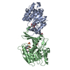
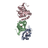
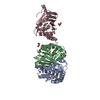
[English] 日本語
Yorodumi
- PDB-3llm: Crystal Structure Analysis of a RNA Helicase -

+
Open data


ID or keywords:

Loading...

-
Basic information

Entry
Database: PDB / ID: 3llm
TitleCrystal Structure Analysis of a RNA Helicase
ComponentsATP-dependent RNA helicase A
KeywordsHYDROLASE / alpha-beta-alpha / Structural Genomics / Structural Genomics Consortium / SGC / Activator / ATP-binding / DNA-binding / Helicase / Methylation / Nucleotide-binding / Nucleus / Phosphoprotein / RNA-binding
Function / homology
Function and homology information


single-stranded 3'-5' DNA helicase activity / positive regulation of viral translation / 3'-5' DNA/RNA helicase activity / CRD-mediated mRNA stability complex / regulation of cytoplasmic translation / regulatory region RNA binding / : / positive regulation of RNA export from nucleus / DNA-templated viral transcription / positive regulation of viral transcription ...single-stranded 3'-5' DNA helicase activity / positive regulation of viral translation / 3'-5' DNA/RNA helicase activity / CRD-mediated mRNA stability complex / regulation of cytoplasmic translation / regulatory region RNA binding / : / positive regulation of RNA export from nucleus / DNA-templated viral transcription / positive regulation of viral transcription / RISC complex binding / triplex DNA binding / positive regulation of DNA topoisomerase (ATP-hydrolyzing) activity / negative regulation of nuclear-transcribed mRNA catabolic process, deadenylation-dependent decay / CRD-mediated mRNA stabilization / protein localization to cytoplasmic stress granule / G-quadruplex DNA unwinding / nucleoside triphosphate diphosphatase activity / nuclear stress granule / 3'-5' RNA helicase activity / perichromatin fibrils / DEx/H-box helicases activate type I IFN and inflammatory cytokines production / RNA secondary structure unwinding / positive regulation of interleukin-18 production / alternative mRNA splicing, via spliceosome / RISC-loading complex / regulation of mRNA processing / miRNA-mediated post-transcriptional gene silencing / RISC complex assembly / regulation of defense response to virus by host / positive regulation of cytoplasmic translation / importin-alpha family protein binding / siRNA binding / RIP-mediated NFkB activation via ZBP1 / positive regulation of response to cytokine stimulus / RNA stem-loop binding / positive regulation of innate immune response / RISC complex / sequence-specific mRNA binding / RNA polymerase binding / 3'-5' DNA helicase activity / DNA duplex unwinding / RNA polymerase II complex binding / cellular response to exogenous dsRNA / DNA replication origin binding / pyroptotic inflammatory response / positive regulation of interferon-alpha production / mRNA transport / positive regulation of DNA repair / DNA helicase activity / positive regulation of interferon-beta production / ribonucleoside triphosphate phosphatase activity / mRNA Splicing - Major Pathway / positive regulation of DNA replication / promoter-specific chromatin binding / DNA-templated transcription termination / transcription coregulator activity / PKR-mediated signaling / cytoplasmic ribonucleoprotein granule / chromatin DNA binding / osteoblast differentiation / positive regulation of inflammatory response / positive regulation of interleukin-6 production / rhythmic process / positive regulation of fibroblast proliferation / ribosome binding / actin cytoskeleton / positive regulation of tumor necrosis factor production / single-stranded DNA binding / double-stranded RNA binding / cellular response to tumor necrosis factor / positive regulation of NF-kappaB transcription factor activity / double-stranded DNA binding / protein-containing complex assembly / RNA polymerase II-specific DNA-binding transcription factor binding / DNA replication / RNA helicase activity / single-stranded RNA binding / transcription coactivator activity / nuclear body / RNA helicase / inflammatory response / ribonucleoprotein complex / RNA polymerase II cis-regulatory region sequence-specific DNA binding / mRNA binding / innate immune response / centrosome / nucleolus / regulation of transcription by RNA polymerase II / ATP hydrolysis activity / positive regulation of transcription by RNA polymerase II / protein-containing complex / DNA binding / RNA binding / nucleoplasm / ATP binding / membrane / metal ion binding / nucleus / cytosol
Similarity search - Function
DHX9, first double-stranded RNA binding domain / DHX9, second double-stranded RNA binding domain / DHX9, DEXH-box helicase domain / : / Helicase associated domain (HA2), ratchet-like / DEAD-box helicase, OB fold / Oligonucleotide/oligosaccharide-binding (OB)-fold / Helicase-associated domain / Helicase associated domain (HA2), winged-helix / Helicase associated domain (HA2) Add an annotation ...DHX9, first double-stranded RNA binding domain / DHX9, second double-stranded RNA binding domain / DHX9, DEXH-box helicase domain / : / Helicase associated domain (HA2), ratchet-like / DEAD-box helicase, OB fold / Oligonucleotide/oligosaccharide-binding (OB)-fold / Helicase-associated domain / Helicase associated domain (HA2), winged-helix / Helicase associated domain (HA2) Add an annotation / Double-stranded RNA binding motif / Double-stranded RNA binding motif / DNA/RNA helicase, ATP-dependent, DEAH-box type, conserved site / DEAH-box subfamily ATP-dependent helicases signature. / Double stranded RNA-binding domain (dsRBD) profile. / Double-stranded RNA-binding domain / DEAD/DEAH box helicase / DEAD/DEAH box helicase domain / Helicase conserved C-terminal domain / helicase superfamily c-terminal domain / Superfamilies 1 and 2 helicase C-terminal domain profile. / Superfamilies 1 and 2 helicase ATP-binding type-1 domain profile. / DEAD-like helicases superfamily / Helicase, C-terminal / Helicase superfamily 1/2, ATP-binding domain / P-loop containing nucleotide triphosphate hydrolases / P-loop containing nucleoside triphosphate hydrolase / Rossmann fold / 3-Layer(aba) Sandwich / Alpha Beta
Similarity search - Domain/homology
ADENOSINE-5'-DIPHOSPHATE / CACODYLATE ION / : / ATP-dependent RNA helicase A
Similarity search - Component
Biological speciesHomo sapiens (human)
MethodX-RAY DIFFRACTION / SYNCHROTRON / MAD / Resolution: 2.8 Å
AuthorsSchutz, P. / Karlberg, T. / Collins, R. / Arrowsmith, C.H. / Berglund, H. / Bountra, C. / Flodin, S. / Flores, A. / Graslund, S. / Hammarstrom, M. ...Schutz, P. / Karlberg, T. / Collins, R. / Arrowsmith, C.H. / Berglund, H. / Bountra, C. / Flodin, S. / Flores, A. / Graslund, S. / Hammarstrom, M. / Johansson, A. / Johansson, I. / Kallas, A. / Kraulis, P. / Kotenyova, T. / Kotzsch, A. / Markova, N. / Moche, M. / Nielsen, T.K. / Nordlund, P. / Nyman, T. / Persson, C. / Roos, A.K. / Siponen, M.I. / Svensson, L. / Thorsell, A.G. / Tresaugues, L. / Van Den Berg, S. / Wahlberg, E. / Weigelt, J. / Welin, M. / Wisniewska, M. / Schuler, H.M. / Structural Genomics Consortium (SGC)
CitationJournal: J.Mol.Biol. / Year: 2010
Title: Crystal structure of human RNA helicase A (DHX9): structural basis for unselective nucleotide base binding in a DEAD-box variant protein.
Authors: Schutz, P. / Wahlberg, E. / Karlberg, T. / Hammarstrom, M. / Collins, R. / Flores, A. / Schuler, H.
History
DepositionJan 29, 2010Deposition site: RCSB / Processing site: RCSB
Revision 1.0May 12, 2010Provider: repository / Type: Initial release
Revision 1.1Jul 13, 2011Group: Version format compliance

-
Structure visualization

Structure viewerMolecule:
MolmilJmol/JSmol

Downloads & links

-
Assembly

Deposited unit
A: ATP-dependent RNA helicase A
B: ATP-dependent RNA helicase A
hetero molecules


Theoretical massNumber of molelcules
Total (without water)54,7258
Polymers53,5322
Non-polymers1,1936
Water48627
1


  • Idetical with deposited unit
  • defined by author&software
TypeNameSymmetry operationNumber
identity operation1_555x,y,z1
Buried area3140 Å2
ΔGint-37 kcal/mol
Surface area21350 Å2
MethodPISA
Unit cell
Length a, b, c (Å)113.700, 113.700, 141.650
Angle α, β, γ (deg.)90.000, 90.000, 120.000
Int Tables number154
Space group name H-MP3221
Noncrystallographic symmetry (NCS)NCS domain:
IDEns-IDDetails
11A
21B

NCS domain segments:
Dom-IDComponent-IDEns-IDRefine codeAuth asym-IDAuth seq-ID
1115A329 - 563
2115B329 - 563

-
Components

-
Protein , 1 types, 2 molecules AB

#1: Protein ATP-dependent RNA helicase A / Nuclear DNA helicase II / NDH II / DEAH box protein 9


Mass: 26765.787 Da / Num. of mol.: 2 / Fragment: Nucleotide binding domain (UNP residues 329-563)
Source method: isolated from a genetically manipulated source
Source: (gene. exp.) Homo sapiens (human) / Gene: DDX9, DHX9, LKP, NDH2 / Plasmid: pNIC-Bsa4 / Production host: Escherichia coli (E. coli) / Strain (production host): BL21(DE3)R3 pRARE
References: UniProt: Q08211, Hydrolases; Acting on acid anhydrides; In phosphorus-containing anhydrides

-
Non-polymers , 5 types, 33 molecules

#2: Chemical ChemComp-MN / MANGANESE (II) ION


Mass: 54.938 Da / Num. of mol.: 2 / Source method: obtained synthetically / Formula: Mn
#3: Chemical ChemComp-CAC / CACODYLATE ION / dimethylarsinate / Cacodylic acid


Mass: 136.989 Da / Num. of mol.: 1 / Source method: obtained synthetically / Formula: C2H6AsO2
#4: Chemical ChemComp-ADP / ADENOSINE-5'-DIPHOSPHATE / Adenosine diphosphate


Mass: 427.201 Da / Num. of mol.: 2 / Source method: obtained synthetically / Formula: C10H15N5O10P2 / Comment: ADP, energy-carrying molecule*YM
#5: Chemical ChemComp-GOL / GLYCEROL / GLYCERIN / PROPANE-1,2,3-TRIOL / Glycerol


Mass: 92.094 Da / Num. of mol.: 1 / Source method: obtained synthetically / Formula: C3H8O3
#6: Water ChemComp-HOH / water / Water


Mass: 18.015 Da / Num. of mol.: 27 / Source method: isolated from a natural source / Formula: H2O

-
Experimental details

-
Experiment

ExperimentMethod: X-RAY DIFFRACTION / Number of used crystals: 1

-
Sample preparation

CrystalDensity Matthews: 4.94 Å3/Da / Density % sol: 75.09 %
Crystal growTemperature: 277 K / Method: vapor diffusion, sitting drop / pH: 6.5
Details: 0.08 M Sodium cacodylate, 0.16 M Calcium acetate hydrate, 14.4 % PEG 8000, 20 % glycerol, pH 6.5, VAPOR DIFFUSION, SITTING DROP, temperature 277K

-
Data collection

DiffractionMean temperature: 100 K
Diffraction sourceSource: SYNCHROTRON / Site: Diamond / Beamline: I03 / Wavelength: 0.98000, 0.98020, 0.96860
DetectorType: ADSC QUANTUM 315 / Detector: CCD / Date: Oct 23, 2009
RadiationProtocol: MAD / Monochromatic (M) / Laue (L): M / Scattering type: x-ray
Radiation wavelength
IDWavelength (Å)Relative weight
10.981
20.98021
30.96861
ReflectionResolution: 2.8→98.51 Å / Num. all: 26628 / Num. obs: 26628 / % possible obs: 100 % / Redundancy: 10.8 % / Rsym value: 0.099 / Net I/σ(I): 14.1
Reflection shellResolution: 2.8→2.95 Å / Redundancy: 11.1 % / Rmerge(I) obs: 0.01 / Mean I/σ(I) obs: 0.7 / Num. measured all: 42589 / Num. unique all: 3834 / Rsym value: 1.041 / % possible all: 100

-
Processing

Software
NameVersionClassificationNB
SCALA3.3.15data scaling
REFMAC5.5.0102refinement
PDB_EXTRACT3.005data extraction
GDAdata collection
MOSFLMdata reduction
SHARPphasing
RefinementMethod to determine structure: MAD / Resolution: 2.8→98.51 Å / Cor.coef. Fo:Fc: 0.944 / Cor.coef. Fo:Fc free: 0.929 / Occupancy max: 1 / Occupancy min: 1 / SU B: 9.989 / SU ML: 0.197 / Cross valid method: THROUGHOUT / σ(F): 0 / ESU R: 0.368 / ESU R Free: 0.263 / Stereochemistry target values: MAXIMUM LIKELIHOOD
Details: HYDROGENS HAVE BEEN ADDED IN THE RIDING POSITIONS. U VALUES: REFINED INDIVIDUALLY
RfactorNum. reflection% reflectionSelection details
Rfree0.238 1343 5 %RANDOM
Rwork0.211 ---
obs0.212 26598 99.95 %-
Solvent computationIon probe radii: 0.8 Å / Shrinkage radii: 0.8 Å / VDW probe radii: 1.4 Å / Solvent model: MASK
Displacement parametersBiso max: 165.68 Å2 / Biso mean: 65.358 Å2 / Biso min: 42.18 Å2
Baniso -1Baniso -2Baniso -3
1-0.82 Å20.41 Å20 Å2
2--0.82 Å20 Å2
3----1.23 Å2
Refinement stepCycle: LAST / Resolution: 2.8→98.51 Å
ProteinNucleic acidLigandSolventTotal
Num. atoms3679 0 67 27 3773
Refine LS restraints
Refine-IDTypeDev idealDev ideal targetNumber
X-RAY DIFFRACTIONr_bond_refined_d0.0150.0223826
X-RAY DIFFRACTIONr_bond_other_d0.0010.022556
X-RAY DIFFRACTIONr_angle_refined_deg1.4571.9835216
X-RAY DIFFRACTIONr_angle_other_deg0.88436251
X-RAY DIFFRACTIONr_dihedral_angle_1_deg5.6115468
X-RAY DIFFRACTIONr_dihedral_angle_2_deg38.26324.379169
X-RAY DIFFRACTIONr_dihedral_angle_3_deg16.38715635
X-RAY DIFFRACTIONr_dihedral_angle_4_deg19.7331526
X-RAY DIFFRACTIONr_chiral_restr0.0730.2605
X-RAY DIFFRACTIONr_gen_planes_refined0.0060.0214180
X-RAY DIFFRACTIONr_gen_planes_other0.0010.02742
X-RAY DIFFRACTIONr_mcbond_it0.6591.52356
X-RAY DIFFRACTIONr_mcbond_other0.1131.5930
X-RAY DIFFRACTIONr_mcangle_it1.32623855
X-RAY DIFFRACTIONr_scbond_it2.03331470
X-RAY DIFFRACTIONr_scangle_it3.4284.51361
Refine LS restraints NCS

Dom-ID: 1 / Auth asym-ID: A / Ens-ID: 1 / Refine-ID: X-RAY DIFFRACTION

NumberTypeRms dev position (Å)Weight position
1385MEDIUM POSITIONAL0.430.5
1672LOOSE POSITIONAL0.715
1385MEDIUM THERMAL0.632
1672LOOSE THERMAL0.710
LS refinement shellResolution: 2.8→2.873 Å / Total num. of bins used: 20
RfactorNum. reflection% reflection
Rfree0.423 82 -
Rwork0.395 1844 -
all-1926 -
obs--100 %

+
About Yorodumi

-
News

-
Feb 9, 2022. New format data for meta-information of EMDB entries

New format data for meta-information of EMDB entries

  • Version 3 of the EMDB header file is now the official format.
  • The previous official version 1.9 will be removed from the archive.

Related info.:EMDB header

External links:wwPDB to switch to version 3 of the EMDB data model

-
Aug 12, 2020. Covid-19 info

Covid-19 info

URL: https://pdbj.org/emnavi/covid19.php

New page: Covid-19 featured information page in EM Navigator.

Related info.:Covid-19 info / Mar 5, 2020. Novel coronavirus structure data

+
Mar 5, 2020. Novel coronavirus structure data

Novel coronavirus structure data

Related info.:Yorodumi Speices / Aug 12, 2020. Covid-19 info

External links:COVID-19 featured content - PDBj / Molecule of the Month (242):Coronavirus Proteases

+
Jan 31, 2019. EMDB accession codes are about to change! (news from PDBe EMDB page)

EMDB accession codes are about to change! (news from PDBe EMDB page)

  • The allocation of 4 digits for EMDB accession codes will soon come to an end. Whilst these codes will remain in use, new EMDB accession codes will include an additional digit and will expand incrementally as the available range of codes is exhausted. The current 4-digit format prefixed with “EMD-” (i.e. EMD-XXXX) will advance to a 5-digit format (i.e. EMD-XXXXX), and so on. It is currently estimated that the 4-digit codes will be depleted around Spring 2019, at which point the 5-digit format will come into force.
  • The EM Navigator/Yorodumi systems omit the EMD- prefix.

Related info.:Q: What is EMD? / ID/Accession-code notation in Yorodumi/EM Navigator

External links:EMDB Accession Codes are Changing Soon! / Contact to PDBj

+
Jul 12, 2017. Major update of PDB

Major update of PDB

  • wwPDB released updated PDB data conforming to the new PDBx/mmCIF dictionary.
  • This is a major update changing the version number from 4 to 5, and with Remediation, in which all the entries are updated.
  • In this update, many items about electron microscopy experimental information are reorganized (e.g. em_software).
  • Now, EM Navigator and Yorodumi are based on the updated data.

External links:wwPDB Remediation / Enriched Model Files Conforming to OneDep Data Standards Now Available in the PDB FTP Archive

-
Yorodumi

Thousand views of thousand structures

  • Yorodumi is a browser for structure data from EMDB, PDB, SASBDB, etc.
  • This page is also the successor to EM Navigator detail page, and also detail information page/front-end page for Omokage search.
  • The word "yorodu" (or yorozu) is an old Japanese word meaning "ten thousand". "mi" (miru) is to see.

Related info.:EMDB / PDB / SASBDB / Comparison of 3 databanks / Yorodumi Search / Aug 31, 2016. New EM Navigator & Yorodumi / Yorodumi Papers / Jmol/JSmol / Function and homology information / Changes in new EM Navigator and Yorodumi

Read more